Dr A Röschl's picture

Second-degree AV Block, Mobitz Type II

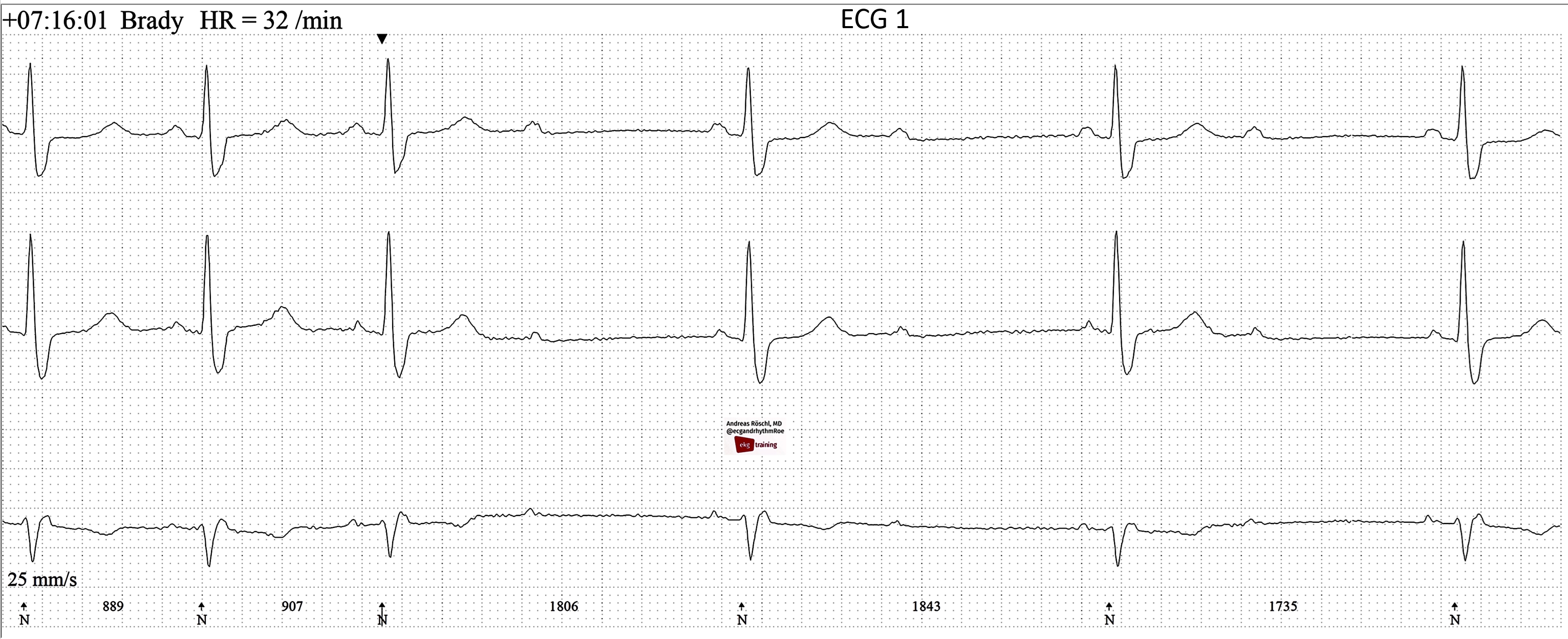
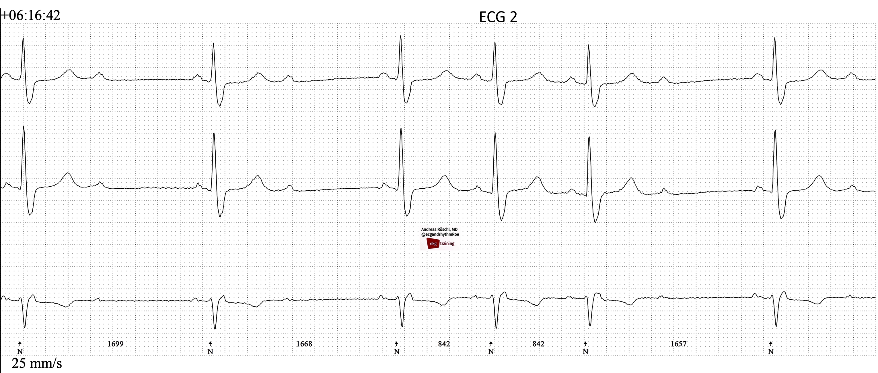
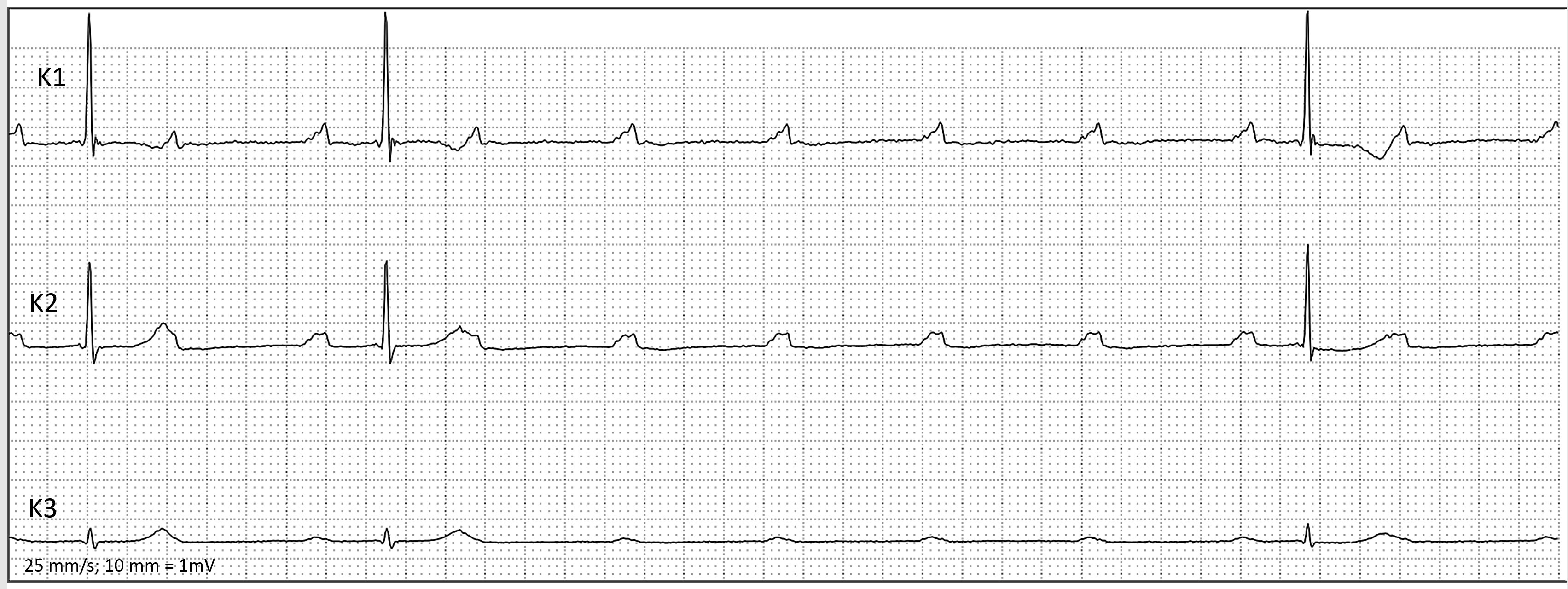
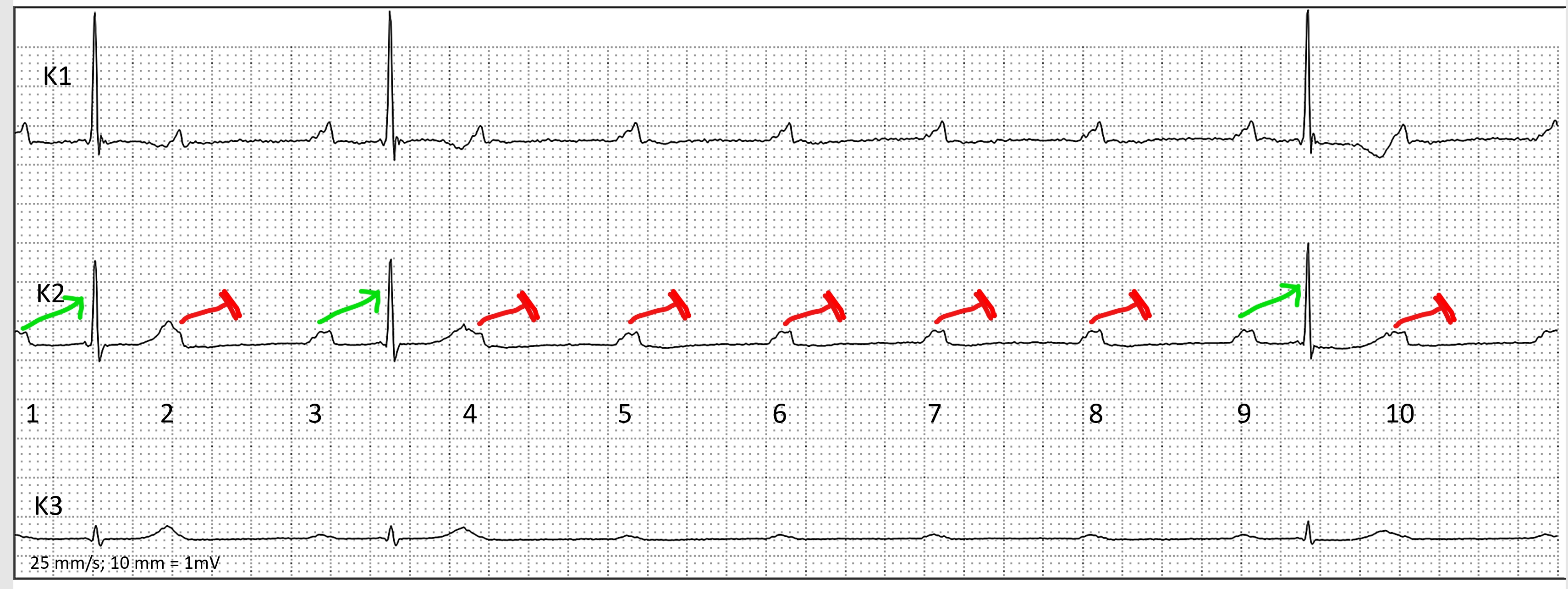
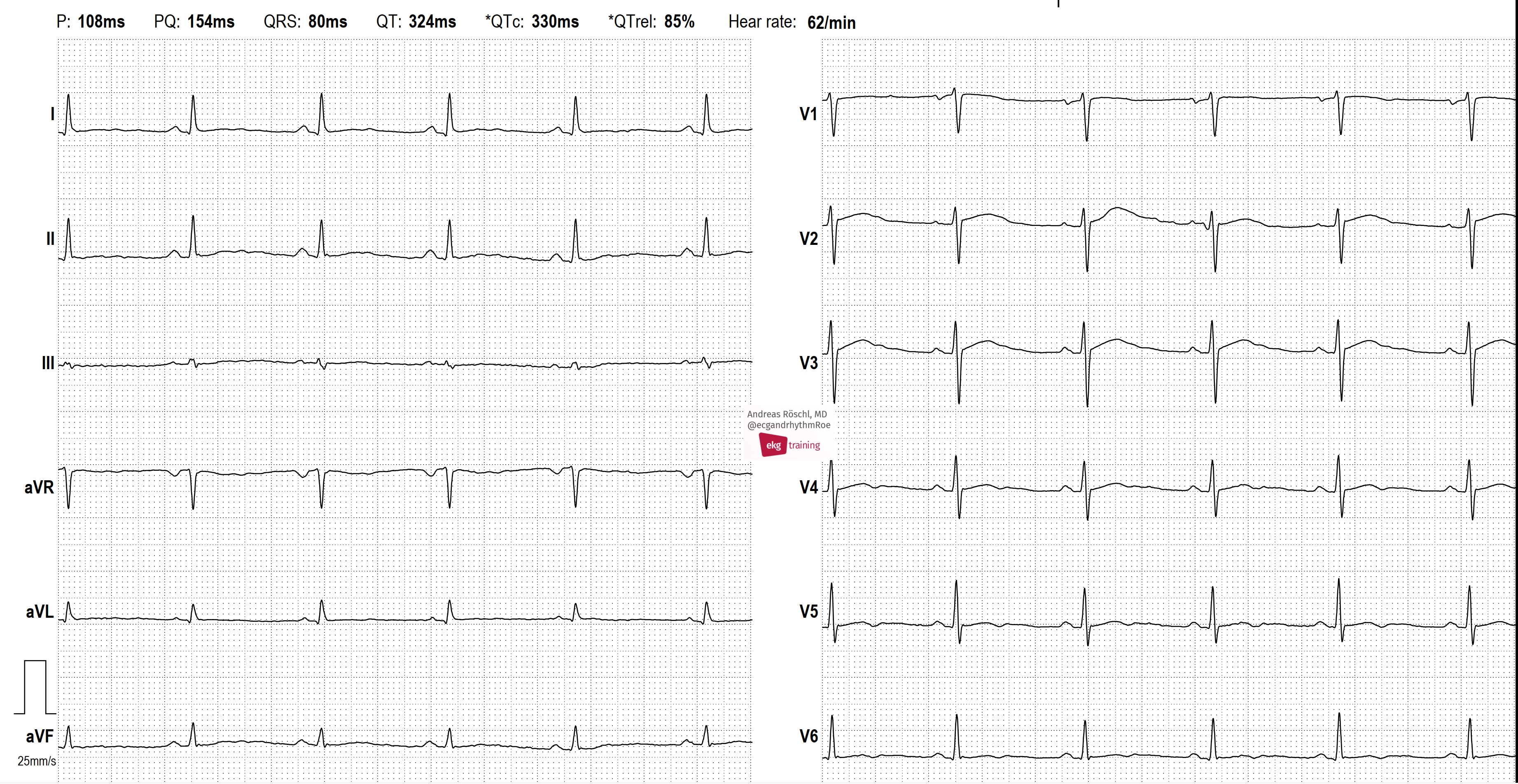
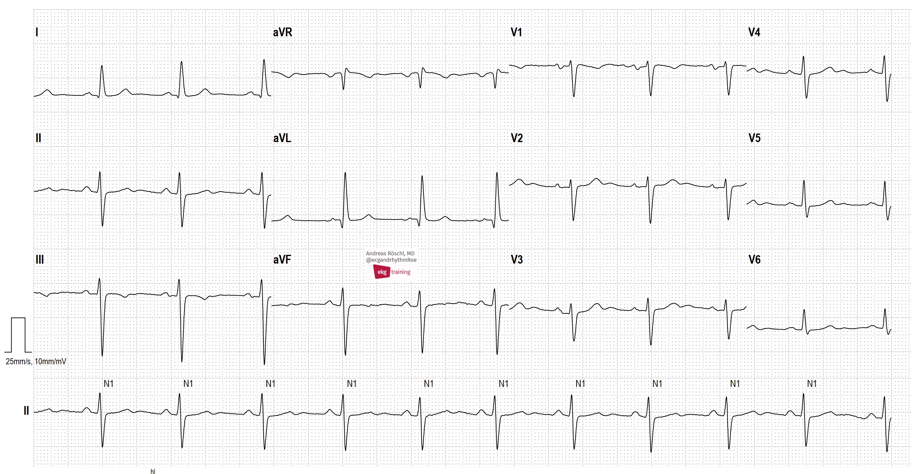
We are observing EKG strip 1 in a Holter EKG recording; what can be said about it? There is a sinus rhythm with a normal PQ interval. After 3 sinus beats, a 2:1 AV block develops. When 2:1 AV block occurs, we should not refer to this as Wenckebach (Mobitz I) or Mobitz II, but rather as a high-grade AV block (other forms include: 3:1, 4:1, 5:1, etc.). The 2:1 block can be intranodally localized and behave benignly like a Wenckebach block typically does. However, it could also be infranodally localized with a potentially serious prognosis.

Dr A Röschl's picture

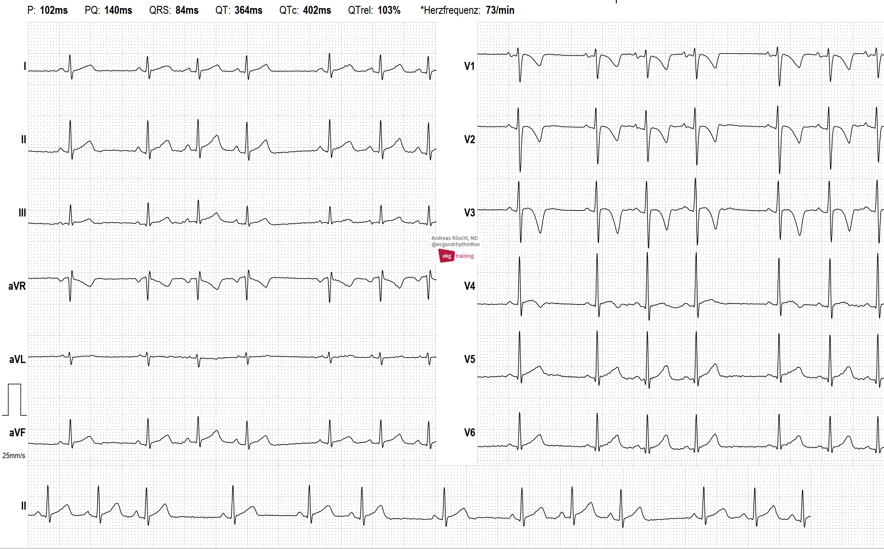
BRUGADA PATTERN

This patient is a 50-year-old man. His brother was diagnosed with epilepsy and died during a seizure at the age of 21.

Parts of the following text I have taken over from Dawn`s explanation on Brugada pattern/Brugada syndrome:

Dr A Röschl's picture

Ventricular Tachycardia In A Patient With CAD And An Old Inferior M.I.

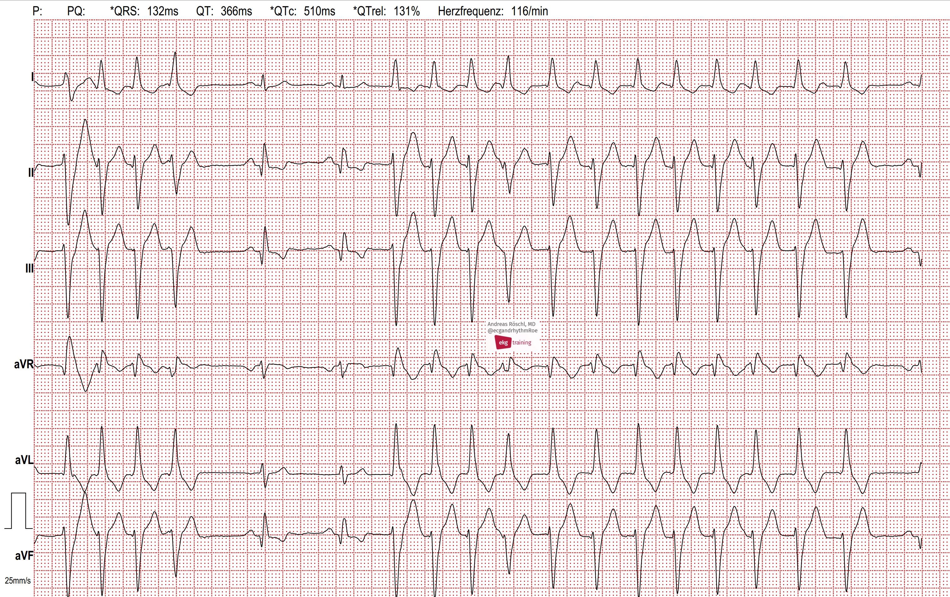
Broad complex tachycardia in a 78-year-old patient with coronary heart disease (CHD) and an old inferior myocardial infarction. Why is this a ventricular tachycardia (VT) and not a supraventricular tachycardia (SVT) with aberrant conduction? Broad complex tachycardia is generally about 80% likely to be ventricular in origin. However, in a patient with CHD and a history of myocardial infarction, this likelihood increases to about 90%. What other clues are there? The tachycardia initiates with a premature QRS complex without a preceding/premature P-wave.

Dr A Röschl's picture

ECG OF A 3 YO BOY, IS IT NORMAL?

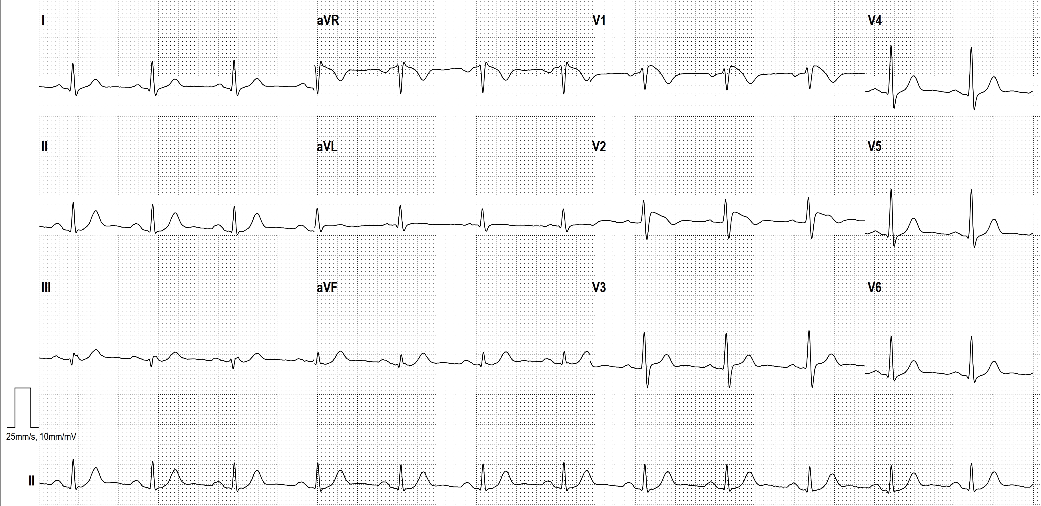
Here is the ECG of a 3-year-old boy. Is there cause for concern? The ECG shows a sinus rhythm with significant sinus arrhythmia. The heart rate increases with inspiration and decreases with expiration, which is called respiratory sinus arrhythmia. The QRS-axis is between 60 and 90 degrees, which is physiological at this age. The negative T-waves in V1-V3 (V4) are also age-appropriately normal. Therefore, there is no reason for concern; the ECG is considered normal for the child's age.

Dr A Röschl's picture

HIGH GRADE AV-Block

Dr A Röschl's picture

VT or SVT? Here, Both!

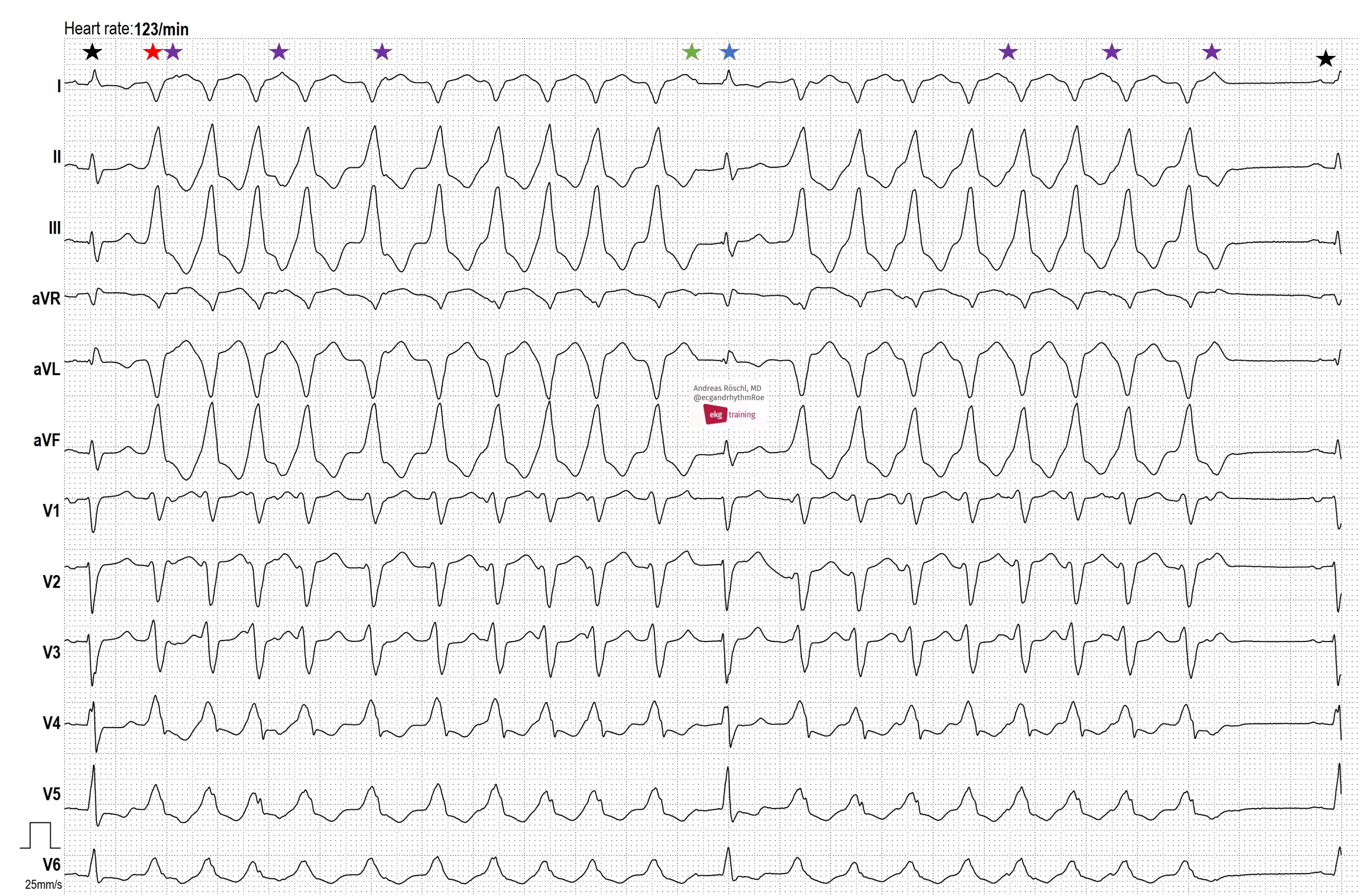
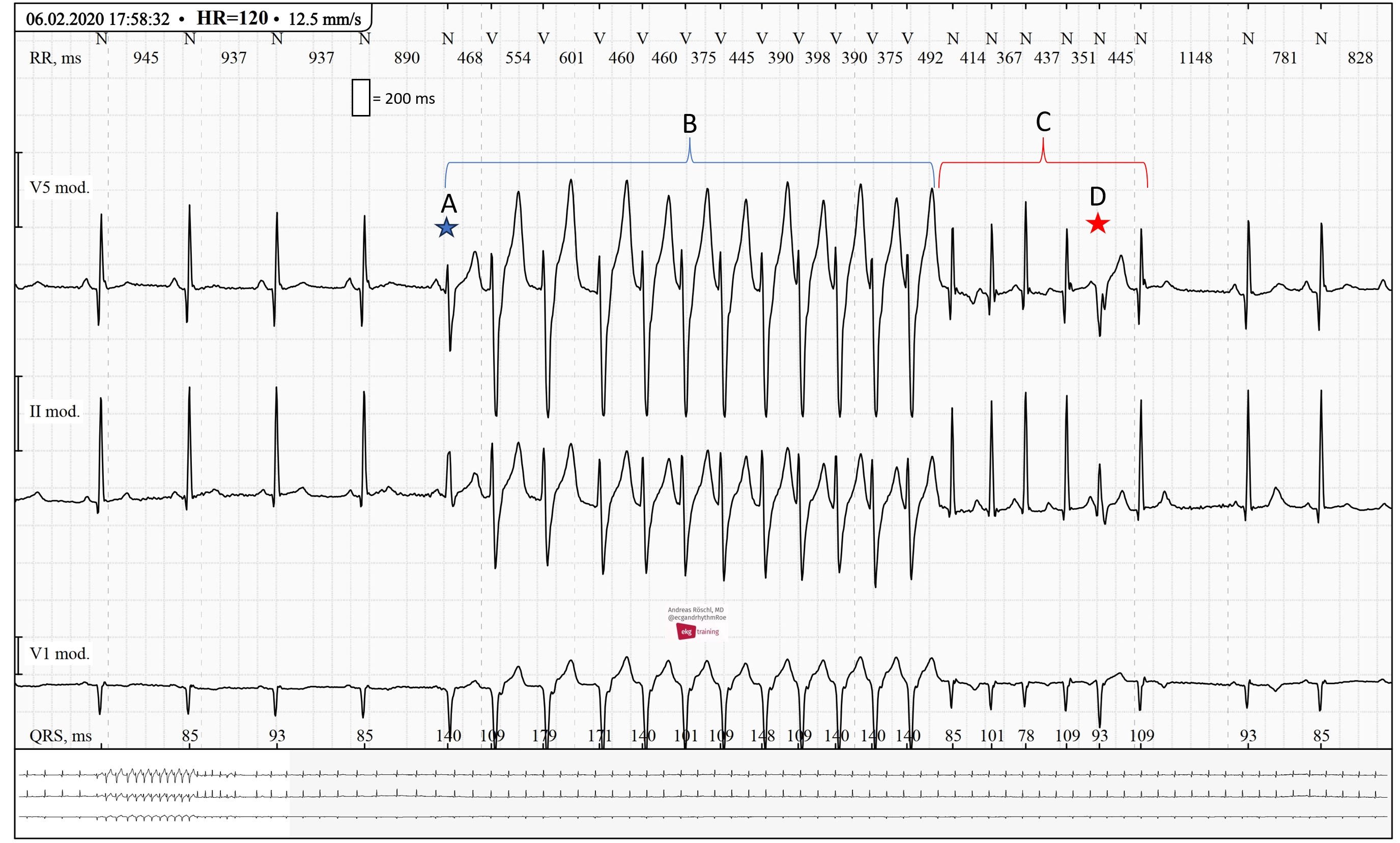
Complex ECGs like this one have to be approached systematically. Firstly, we can see a normal sinus rhythm. A is the first beat of a wide complex tachycardia. This must be a ventricular tachycardia. Although there is a P-wave before the first beat of the tachycardia, it is not premature. Therefore, there is no SVT with aberrant conduction. The first beat of the tachycardia looks different from the subsequent beats because there is a fusion beat present. The VT conducts 1:1 back to the atria (a small negative P-wave can be seen at the end of the QRS complex in V1).

Dr A Röschl's picture

Long QT Syndrome,Look Closely and Do Not Trust the ECG-Computer

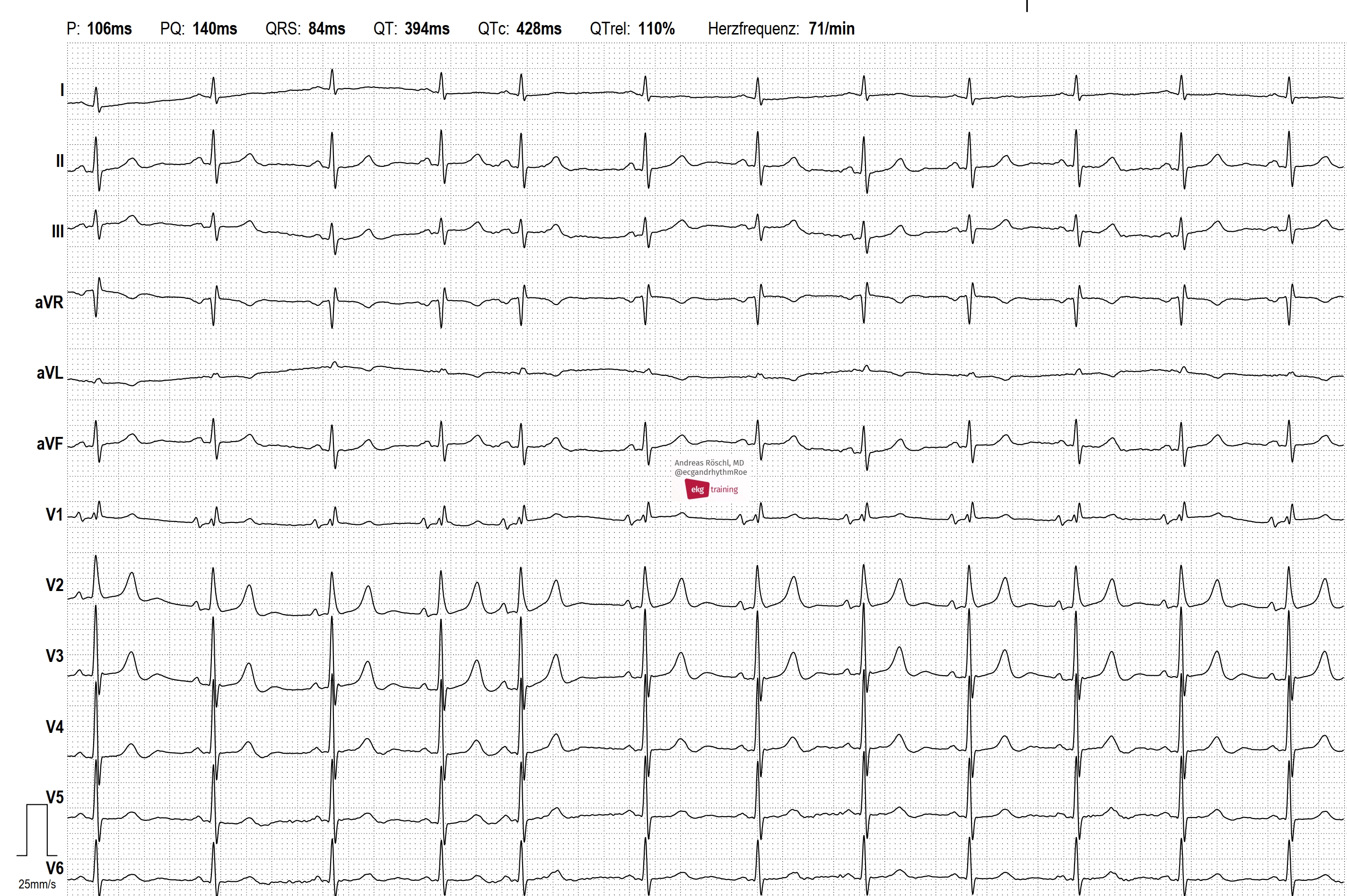
ST-elevation V2/V3, why? The patient never had chest pain, echocardiography inconspicious. History of several syncopal episodes. But, there is something wrong with the ECG. We observe a sinus rhythm ECG, at first glance, there are ST-elevations in leads V2-V4. No ST-depressions are visible. Upon closer examination, a prominent T-wave is noted, starting immediately after the QRS complex. The ECG computer erroneously indicates a significantly shortened QT and QTc interval.

Dr A Röschl's picture

Ventricular Tachycardia After Inferior Myocardial Infarction

76-year-old man, with a history of inferior wall myocardial infarction. He experiences recurrent episodes of brief palpitations, often lasting only 5-15 seconds. In this ECG, at the beginning, the last part of a wide-complex tachycardia is visible. After 2 sinus beats, another wide-complex tachycardia begins (with the same QRS configuration).

Dr A Röschl's picture

Non-respiratory Sinus Arrhythmia

This is the ECG of an 81-year-old man with hypertension. Currently, he has no complaints: no palpitations, no shortness of breath, no syncope, no chest pain. The rhythm is irregular, but all P-waves have the same shape. There is a non-respiratory sinus arrhythmia present, which is essentially the minimal variant of a sick sinus syndrome. There are no consequences for the patient resulting from this diagnosis.

Dr A Röschl's picture

Sudden Shortness of Breath - Pulmonary Embolism

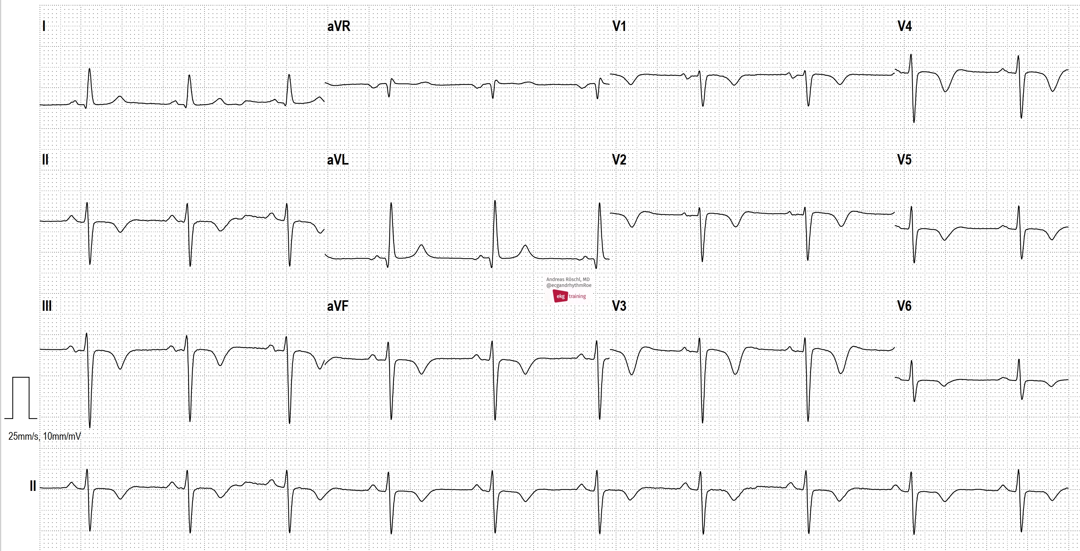
ECG 1 is from a 57-year-old male with no prior cardiac disease. He reports acute shortness of breath for 2 days. We see a sinus rhythm with left anterior fascicular block (LAFB) and conspicuous T-wave inversions in the inferior leads and in V1-V6. These are typical ECG changes that may indicate a pulmonary embolism. ECG 2 was taken from the same patient 1 year earlier. The patient has an acute pulmonary embolism. Sinus tachycardia may be present in acute pulmonary embolism. However, as in this example, the heart rate can also be completely normal.

Pages

All our content is FREE & COPYRIGHT FREE for non-commercial use

Please be courteous and leave any watermark or author attribution on content you reproduce.