
Submitted by Dr A Röschl on Mon, 01/13/2025 - 01:49

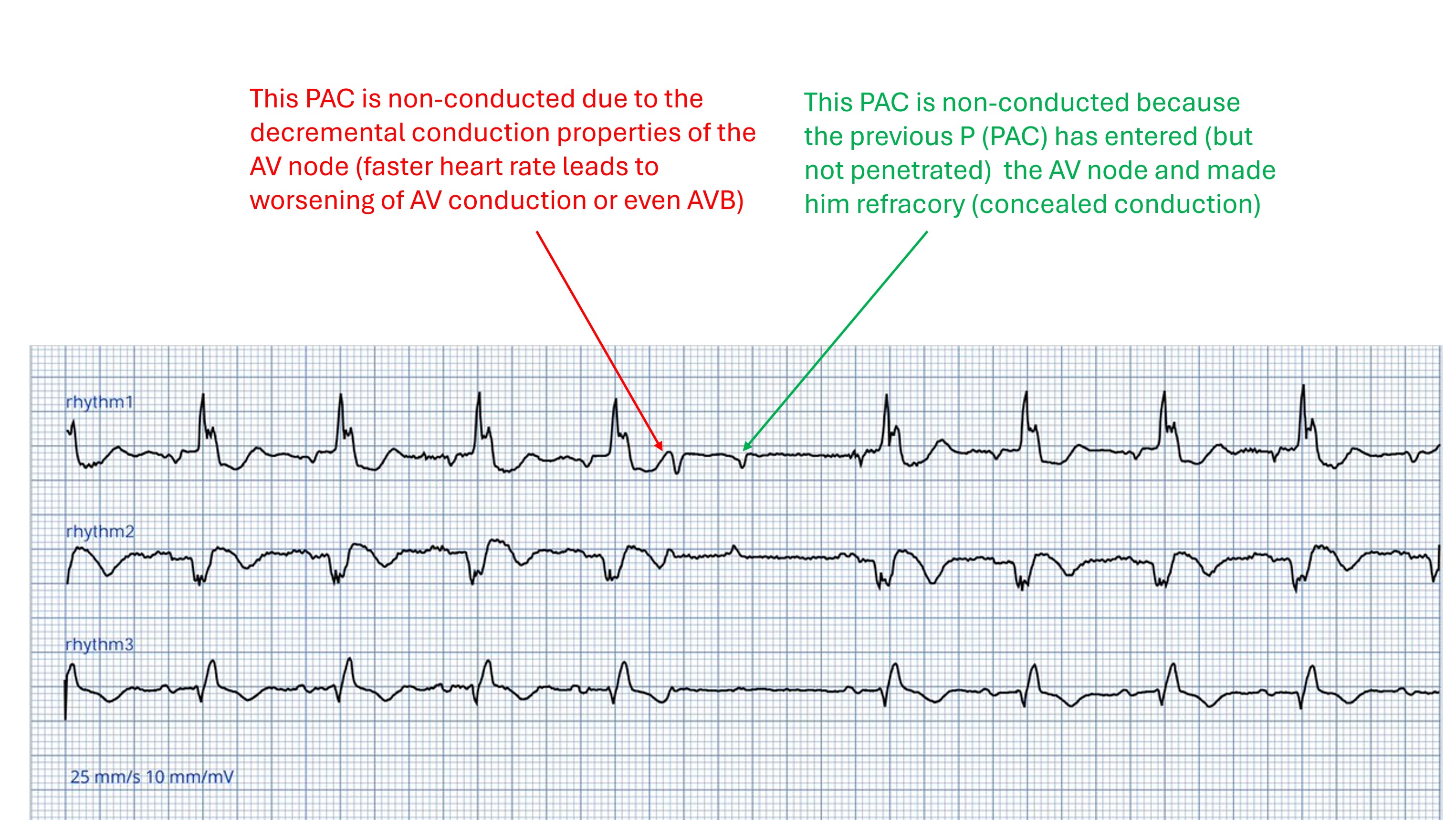
This ECG (3 rhythm strips) initially shows a sinus rhythm with 1st degree AVB grade I and wide QRS complexes (presumably RBBB pattern). A PAC (P-wave premature, different form than in sinus rhythm) appears approximately in the middle of the ECG, this is not conducted . Due to the strong prematurity, this is not surprising. But why is the 2nd PAC also blocked? The answer can be found in the second picture.
Rate this content:
-

- Dr A Röschl's blog
- Log in or register to post comments
All our content is FREE & COPYRIGHT FREE for non-commercial use
Please be courteous and leave any watermark or author attribution on content you reproduce.