
Submitted by Dr A Röschl on Wed, 07/26/2023 - 06:20
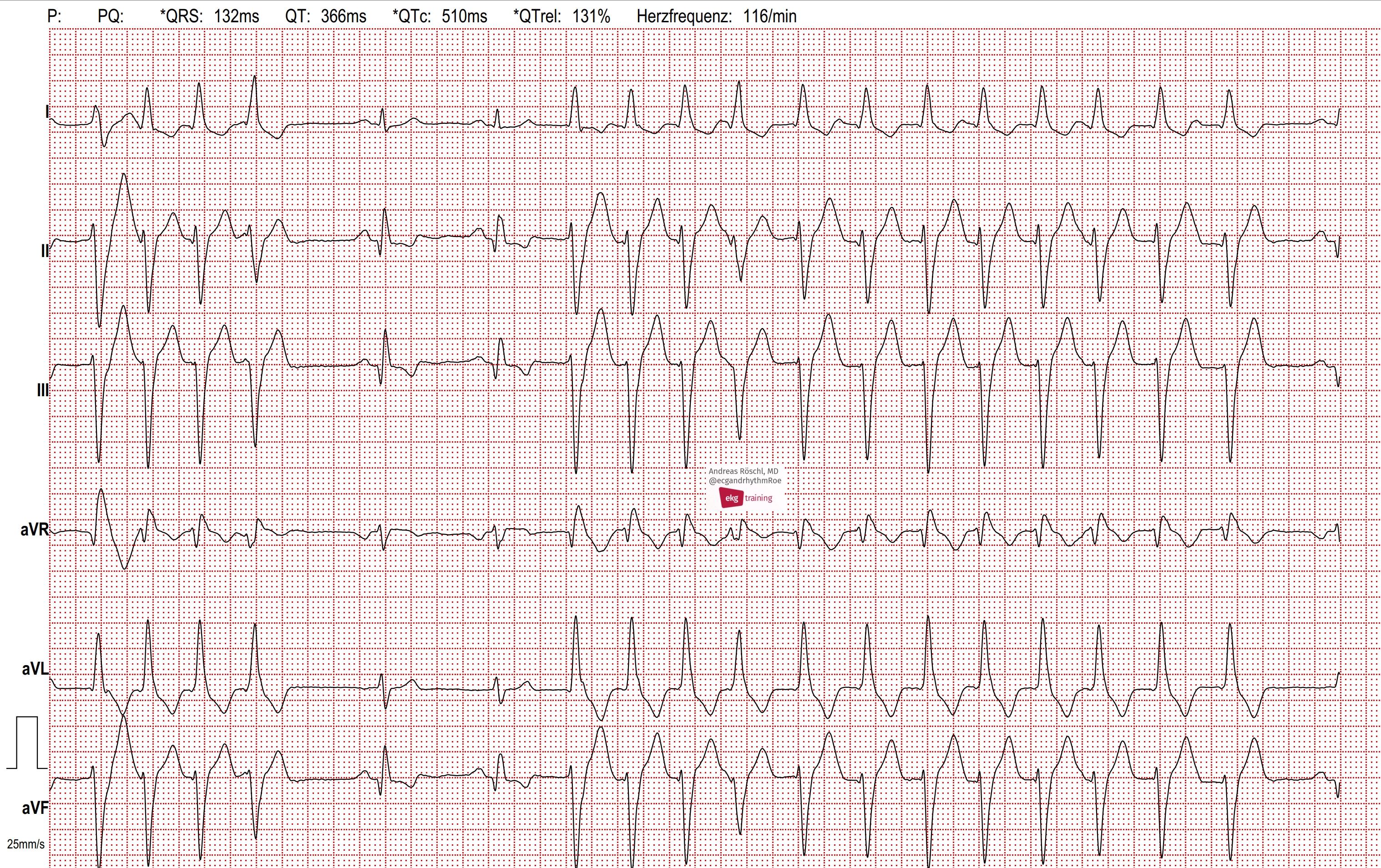
76-year-old man, with a history of inferior wall myocardial infarction. He experiences recurrent episodes of brief palpitations, often lasting only 5-15 seconds. In this ECG, at the beginning, the last part of a wide-complex tachycardia is visible. After 2 sinus beats, another wide-complex tachycardia begins (with the same QRS configuration). Approximately 80% of wide-complex tachycardias are ventricular tachycardias, with the rest being distributed among sinus tachycardias or supraventricular tachycardias with preexisting or functional bundle branch block, and very rarely, antidromic WPW tachycardia. In the present case, there is a history of inferior wall myocardial infarction, which can be verified by the Q waves in the inferior leads II, III, and aVF. Consequently, the probability of ventricular tachycardia (VT) is over 90%. Furthermore, the tachycardia begins with a premature broad QRS complex without a preceding premature p-wave. The 4th beat of the second tachycardia is likely a fusion beat. There is no doubt about the presence of VT in this case.
Rate this content:
-

- Dr A Röschl's blog
- Log in or register to post comments
All our content is FREE & COPYRIGHT FREE for non-commercial use
Please be courteous and leave any watermark or author attribution on content you reproduce.
Comments
Excellent Discussions
Dr. Röschl...
I enjoy your discussions very much and always look forward to reading them.
Jerry W. Jones, MD
Jerry W. Jones MD FACEP FAAEM
https://www.medicusofhouston.com
Twitter: @jwjmd