Displaying 1 - 3 of 3
Dawn's picture

Instructors' Collection ECG: Anterior-lateral M.I.

The Patient: This ECG was obtained from a man in his mid-sixties who was complaining of chest pain.  The pain had an acute onset and is described as "10" on a 1-10 scale.  He has a PMHx of coronary artery disease with stents in his right coronary artery and minimally invasive aortic valve replacement.

The ECG:  The rate is 86 bpm.  The rhythm is normal sinus rhythm with one PAC (10th beat).  The PR interval is .18 seconds (176 ms), the QRS duration is .122 seconds (.12 seconds). This represents a ventricular conduction delay. There is no right or left bundle branch block.   The QT/QTc is 333 ms/400 ms (B). The frontal plane QRS axis is leftward, with criteria for left anterior fascicular block. LAFB can be explained by this patient's history of prior CAD and valve replacement.  There is ST elevation in Lead I and also in V1-V6.  The ST segments have a straight shape in Leads I and aVL and in V1-V6.  This shape represents ischemia in a patient with these symptoms and ECG findings.  This is an ANTERIOR-LATERAL OCCLUSIVE M.I.

Followup:   The patient was taken to the cath lab and had angioplasty of an occlusive mid-LAD (left anterior descending) lesion and a partially-occlusive mid-RCA lesion.  

Dawn's picture

Myocardial Infarction With Non-obstructive Coronary Arteries

This ECG was obtained from a 37-year-old male who was complaining of non-radiating substernal chest pain.  He offered no significant medical history.  He denied taking any medications.  He was hypertensive and bradycardic on arrival in the Emergency Dept. He was alert and ambulatory.  Approximately 20 minutes after first being seen by paramedics, he suffered an episode of ventricular fibrillation in the E.D.  He was resuscitated and sent to the cath lab.  His coronary arteries were without lesions.  We do not know the results of any lab tests, including troponins.

What does the ECG show?  The rhythm is sinus bradycardia at a rate of 48 bpm. The PR, QTc, intervals and QRS duration are normal.  The QRS frontal plane axis is normal and there is good R wave progression in the precordial leads.  There is ST segment elevation in Leads I, aVL, V2, and V3, with reciprocal ST depression in Leads III and aVF.  The ST segments that are elevated retain a relatively “normal” shape, being concave upward. There are no abnormal T wave inversions or pathological Q waves.

Dawn's picture

Acute Anterior-lateral M.I. With Right Bundle Branch Block and Left Posterior Fascicular Block

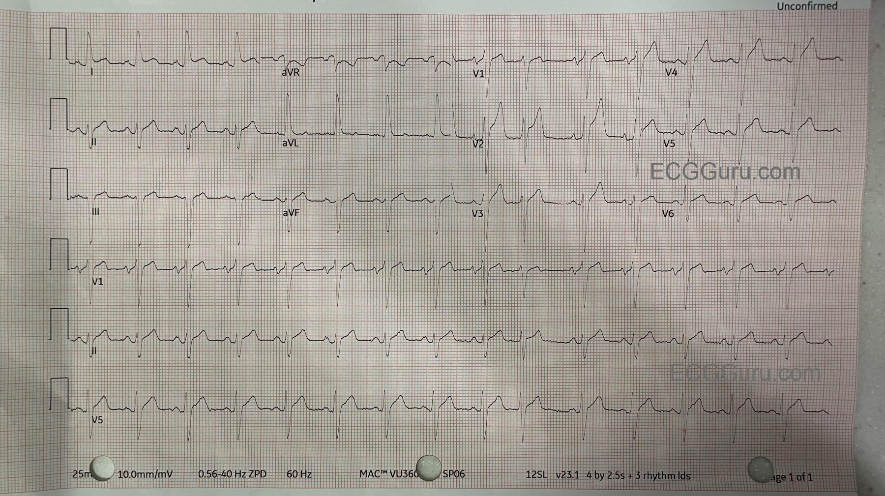
This ECG was obtained from a patient who suffered an occlusion of the left main coronary artery.  ST elevation is seen in Leads V1 through V6, as well as I and aVL.  This is an indicator that the circumflex artery is included in this M.I., and the occlusion is above the bifurcation of the LM and the circ.  The patient also has a right bundle branch block and a left posterior fascicular block.  This bi-fascicular block can be a dangerous complication of acute M.I., as two of the three main bundle branches are no longer functional.

The ECG shows typical ST depression, probably reciprocal to the elevation, in the inferior leads.

The right bundle branch block is diagnosed by the following criteria:  1) Wide QRS;  2) Supraventricular rhythm; and 3) rSR' pattern in V1 with Rs with a wide little s wave in Leads I and V6.

The left posterior fascicular block is diagnosed by right axis deviation and by ruling out other causes of right axis deviation.  In RAD, Lead III will have a taller positive ( R ) wave than Lead II, and a negative Lead I.

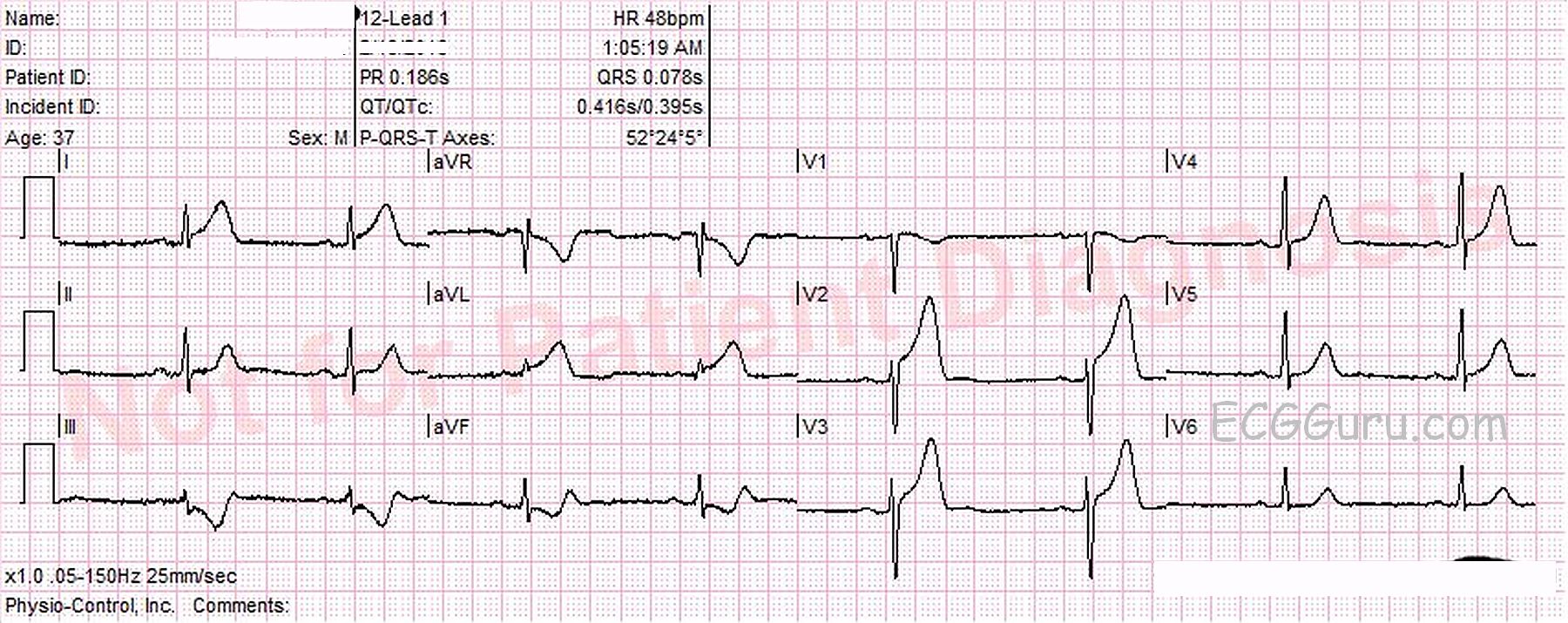
This type of occlusion is often called the "Widow Maker", and requires very rapid intervention to restore blood flow and prevent complicatons.  If there is good news, it is that there are no pathological Q waves, which would indicate necrosis, and this patient was taken quickly to a full-service cardiac center with interventional cath labs and open heart surgery available.

All our content is FREE & COPYRIGHT FREE for non-commercial use

Please be courteous and leave any watermark or author attribution on content you reproduce.