The Patient:A 64-year-old man complaining of chest pain and shortness of breath for 20 minutes.Long-standing history of triple vessel disease, severe aortic stenosis, hypertension, thrombocytopenia.Meds unknown.He was not considered to be a candidate for valve surgery.
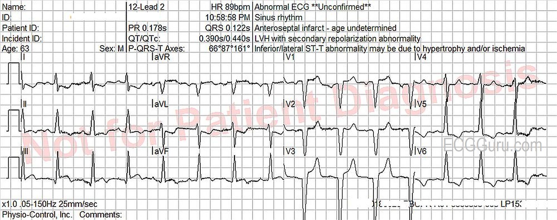
The ECG: There is normal sinus rhythm with a rate of 90 bpm.P waves are not visualized well in all leads, so remember that the three channels of this ECG are run simultaneously.If you see a P wave in Leads I and II, they are also present in Lead III.The PR interval is WNL.
The QRS complexes are wide, at .122 seconds (122 ms).The criteria for left bundle branchblock are met. (Supraventricular rhythm, wide QRS, upright QRS in Leads I and V6, negative QRS in V1).The frontal plane axis is within normal limits, but toward the right, at 87 degrees.The QRS complexes transition at V4 from negative to positive, but Leads V1 – V3 have no initial r waves.These are possibly pathological Q waves, likely from a past anterior-septal M.I.