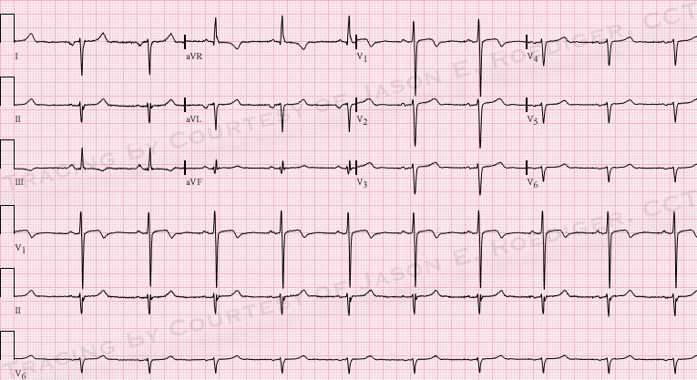
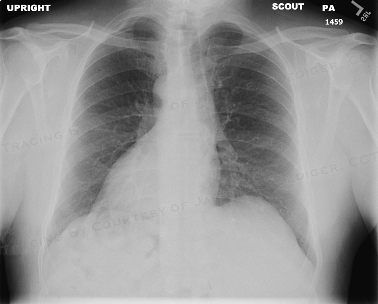
This middle-aged man presents to the Emergency Dept. complaining of shortness of breath and fever. Blood work, chest xray, and ECG are ordered. When the ER Tech obtains the 12-Lead ECG, he is puzzled by the following findings:
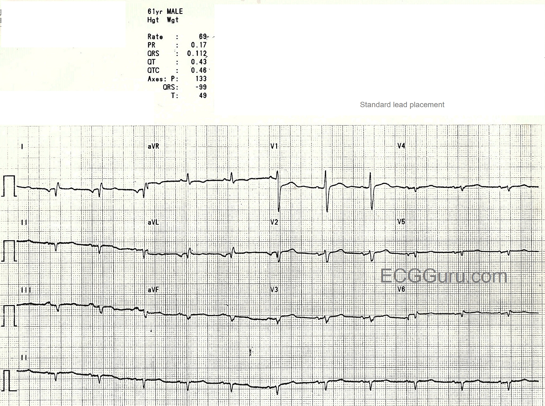
Low voltage in the chest leads V2 - V6
Abnormal right axis of P waves with negative P waves in Leads I and aVL
Abnormal right QRS axis with an upright aVR.
Seeing his puzzled look, the patient says, "Oh, I forgot to tell you, I have mirror-image dextrocardia!"
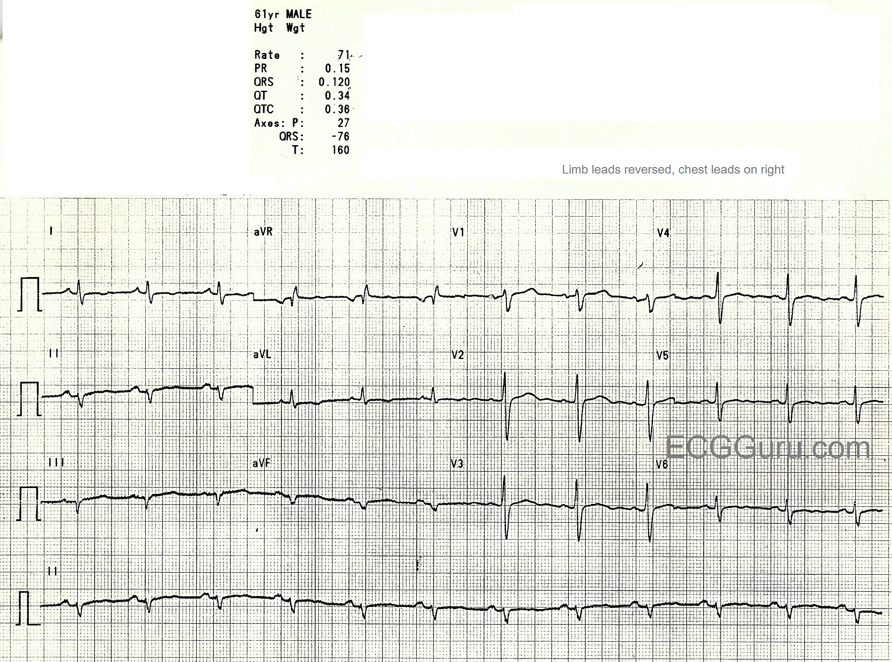
After consulting with the ED physician, the Tech reverses the arm leads and the chest leads and obtains the second ECG.
The P wave axis is now correct, although there is still derangement of the QRS axis. The QRS complexes in the chest leads look more normal, but do not show the usual R wave progression of the normal ECG.