
Submitted by Dr A Röschl on Mon, 07/31/2023 - 03:59
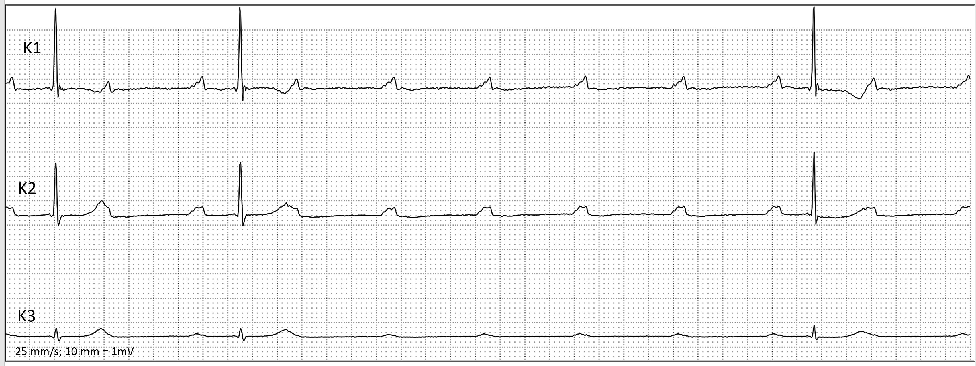
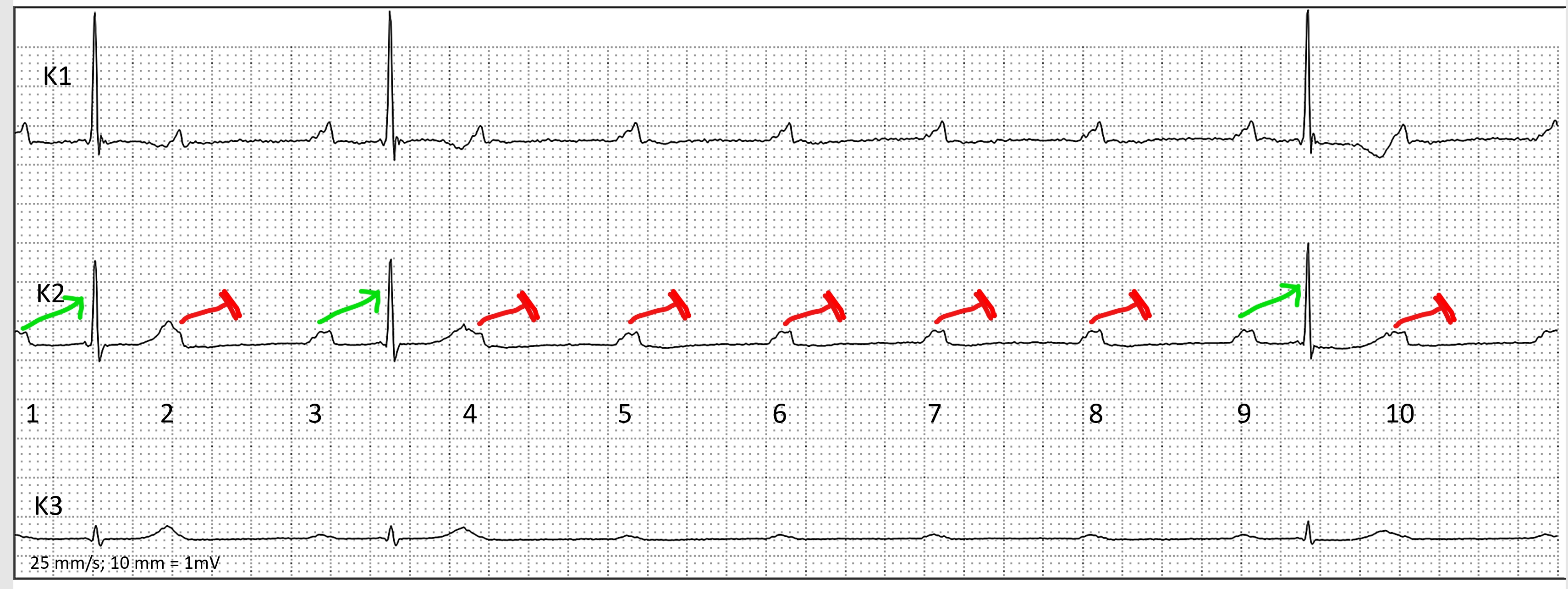
Here you can see the ECG (not labeled and labeled) of a 78-year-old lady with recurrent syncopal episodes. At point 1, you can observe atrial activity (sinus rhythm) that is transmitted to the ventricles with a prolonged PR interval. The next P wave at point 2 (partially hidden in the T-wave) is blocked, but the P wave after that is conducted again. This indicates a short phase of 2:1 AV block. The P waves 4-8 are all not conducted, and P wave 9 is conducted with a first-degree AV block again, while P wave 10 is blocked.
In the middle of the ECG, there is a 6:1 AV conduction, which is referred to as a high grade AV block. What is the difference from complete heart block (CHB)? In CHB, there is NO AV conduction; atria and ventricles are acitvated independently of each other."
Rate this content:
-

- Dr A Röschl's blog
- Log in or register to post comments
All our content is FREE & COPYRIGHT FREE for non-commercial use
Please be courteous and leave any watermark or author attribution on content you reproduce.