
Submitted by Dr A Röschl on Sun, 07/30/2023 - 02:07
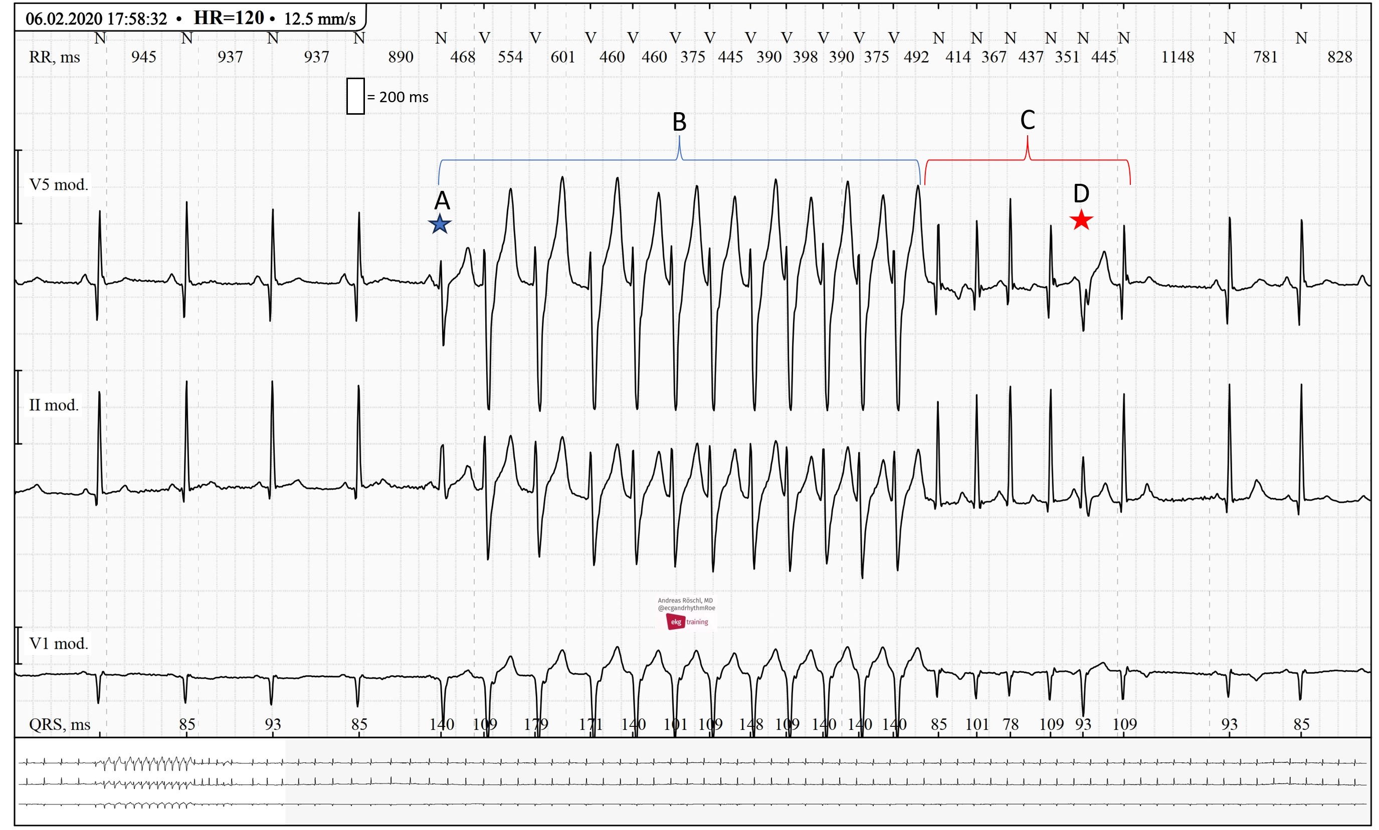
Complex ECGs like this one have to be approached systematically. Firstly, we can see a normal sinus rhythm. A is the first beat of a wide complex tachycardia. This must be a ventricular tachycardia. Although there is a P-wave before the first beat of the tachycardia, it is not premature. Therefore, there is no SVT with aberrant conduction. The first beat of the tachycardia looks different from the subsequent beats because there is a fusion beat present. The VT conducts 1:1 back to the atria (a small negative P-wave can be seen at the end of the QRS complex in V1). At C, the wide-complex tachycardia converts into a narrow-complex tachycardia, indicating that the VT has stopped and an SVT has started (due to V-A conduction). It could be an AVNRT, but this cannot be confirmed with certainty. At D, a PVC is seen within the SVT (which stops after 5 beats). A transition of VT into SVT is rare, but it is essential to know that such a phenomenon exists.
Rate this content:
-

- Dr A Röschl's blog
- Log in or register to post comments
All our content is FREE & COPYRIGHT FREE for non-commercial use
Please be courteous and leave any watermark or author attribution on content you reproduce.