Displaying 1 - 10 of 15
Dr A Röschl's picture

Ventricular Tachycardia After Inferior Myocardial Infarction

76-year-old man, with a history of inferior wall myocardial infarction. He experiences recurrent episodes of brief palpitations, often lasting only 5-15 seconds. In this ECG, at the beginning, the last part of a wide-complex tachycardia is visible. After 2 sinus beats, another wide-complex tachycardia begins (with the same QRS configuration).

Dawn's picture

Wide Complex Tachycardia

This pair of ECGs feature one of our recurring themes:  wide-complex tachycardia (WCT). It is a fascinating topic, as tachycardia has many causes and many mechanisms, and wide QRS also has many causes, with the mechanism being slow conduction through the ventricles. 

Sometimes, it is not possible to diagnose the true origin of a WCT from one ECG, or even serial ECGs.  Is the tachycardia due to increased sympathetic activity (fear, dehydration, exercise, hypoxia, hypovolemia, etc.)?  Or is the fast rate due to reentry, where one impulse gets “caught” in a loop, repeating itself rapidly, and depolarizing the myocardium with each pass?  What is the location of the pacemaker that is responsible for the rhythm?  Is it a supraventricular rhythm that has suffered an intraventricular conduction delay, widening the QRS?  Or is the rhythm originating in a ventricular pacemaker, without the ability to travel on the fast highway that is the intraventricular conduction system? 

If you or your students work in an acute care setting, such as pre-hospital or emergency department, you may not be with the patient long enough or be able to conduct enough tests to determine without a doubt the answers to the above questions.  Some WCTs cause such severe symptoms that they must be dealt with quickly, to avoid rapid deterioration to ventricular fibrillation.  For that reason, there is a widely-accepted rule for WTC treatment:

TREAT ALL WIDE-COMPLEX TACHYCARDIA AS IF IT IS V TACH UNTIL PROVEN OTHERWISE.    

Dawn's picture

Wide Complex Tachycardia

The Patient   A 64-year-old woman has called 911 because she has chest discomfort radiating to her left arm, palpitations, weakness, and a headache.  She had a valve replacement (we do not know which valve) two weeks ago and has a healing incision over her sternum.  She is found sitting in a chair, pale, cool, and diaphoretic. Her blood pressure is 94/palp.  Her pulse rate is 196 bpm and weak. She is afebrile.

ECG #1   This ECG shows a wide-complex tachycardia at 196 bpm.  The QRS complexes are .132 seconds in duration, per the ECG machine. The rate is too fast to appreciate whether there are P waves present.  We did not see the onset of the tachycardia, but with a rate this fast and regular, it is most likely a reentrant rhythm, rather than sinus tachycardia.  An abrupt onset of the rhythm would point to a diagnosis of a reentrant rhythm, either ventricular tachycardia (VT) or paroxysmal supraventricular tachycardia (PSVT). 

There is an important rule in emergency medical care:  a wide-complex tachycardia should be treated as VT until and unless it is proven to be something else.  The most likely alternate interpretation is PSVT with aberrant conduction, which usually takes the form of left or right bundle branch block. Fortunately, the paramedics on this call have a protocol for treating WCT that includes electrical cardioversion for the unstable patient, and amiodarone for the stable patient.  This protocol serves both possibilities, VT and PSVT, well.  The patient’s perfusion status and BP made her borderline in this determination, but she was alert and oriented, so the paramedics opted for administering the amiodarone while they prepared to electrically cardiovert.

Dawn's picture

Wide Complex Tachycardia

The Patient:   The details of this patient’s complaints and presentation are lost, but we know he was a 66-year-old man who was being treated in the Emergency Department. His rhythm went from sinus tachycardia with non-respiratory sinus arrhythmia to multi-focal atrial tachycardia (MAT) to wide-complex tachycardia. The WCT lasted a few minutes and spontaneously converted to an irregular sinus rhythm.

Wide-complex tachycardia:  Ventricular tachycardia or aberrantly-conducted supraventricular tachycardia?  When confronted with a wide-complex tachycardia, it can be very difficult to determine whether the rhythm is ventricular or supraventricular with aberrant conduction, such as bundle branch block. The patient’s history and presentation may offer clues.  It is very important, if the patient’s hemodynamic status is at all compromised (they are “symptomatic”), the WCT should be treated as VENTRICULAR TACHYCARDIA until proven otherwise.  

There have been many lists made of the ECG features that favor a diagnosis of ventricular tachycardia. Here are two such lists:  Life In The Fast Lane, and National Institute of Health.

The ECG:  This ECG shows a regular, fast, wide-QRS rhythm.  The rate is 233 bpm.  It had a sudden onset and sudden offset (not shown on this ECG), and the rhythm lasted about 3-5 minutes. The patient felt the change in rate, but did not become hypotensive or unstable.  Some features that relate directly to the most commonly-referenced VT vs. SVT charts are:

Dawn's picture

Ventricular Tachycardia In A Patient With Myopathy

These two ECGs are from a 77-year-old woman who was complaining of palpitations and mild shortness of breath.  She stated a history of atrial fibrillation.  She was alert, with a systolic BP over 120.  At the hospital, she was found to have cardiomyopathy, resulting in global hypokinesis. She also had significant coronary artery narrowing in her left main, left anterior descending, and circumflex, which were treated with coronary artery bypass graft surgery.

The first ECG was taken on arrival of the EMS crew at the patient’s home.  It shows ventricular tachycardia, rate 226 bpm, All WCTs should be considered to be ventricular tachycardia until proven otherwise.  While WCT can sometimes be difficult to definitively diagnose in the field, this ECG has many features which favor the diagnosis of VT, including:

·         An extremely wide QRS (I measure .24 sec., the machine measures .368 sec.).

·         An extreme left axis deviation (aVF is all negative).

·         Absence of either RBBB or LBBB pattern, with a  completely negative QRS in V6.  This all negative V6 places the liklihood of the rhythm being VT to about 100%.

Dawn's picture

Right Ventricular Outflow Tract Tachycardia (RVOT)

This ECG was taken from a patient who was complaining of palpitations and tachycardia, but who was hemodynamically stable, with no history of heart disease.  It is an example of RIGHT VENTRICULAR OUTFLOW TRACT TACHYCARDIA, a type of idiopathic ventricular tachycardia.  The ECG signs of RVOT are:  wide QRS complex, left bundle branch block pattern (QRS negative in V1 and positive in Leads I and V6), heart rate over 100 bpm, rightward or inferior axis (LBBB usually has a normal to leftward axis), AV dissociation.

RVOT accounts for about 10% of all ventricular tachycardias, and 70% of idiopathic VT.  It is most often found in structurally normal hearts, but it may occur in patients with arrhythmogenic right ventricular dysplasia.  For more on RVOT, read Life In the Fast Lane.

RVOT tachycardia sometimes converts with adenosine.  The patient in this example converted after being administered amiodarone.

Dawn's picture

Wide Complex Tachycardia

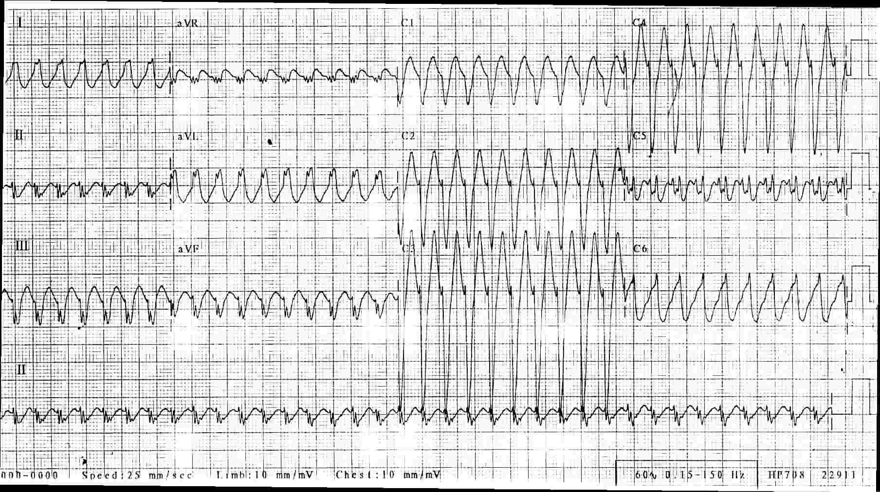
Wide-QRS rhythms can be difficult to diagnose from the ECG alone.  This difficulty is compounded when the rate is fast, as it can be hard to determine if P waves are present before the QRSs, or dissociated, or absent.

This ECG and rhythm strip were donated to the ECG Guru by Ryan Cihowiak.  We don't have clinical information on the patient, unfortunately.  It is a great example, however, of how difficult WCT can be to diagnose.

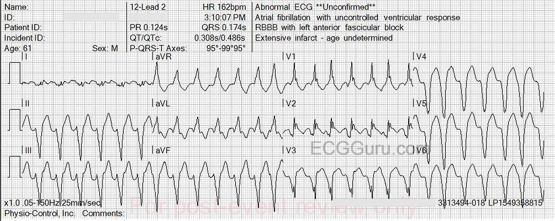
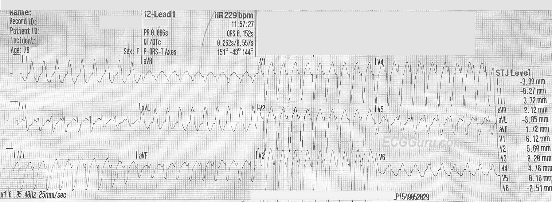
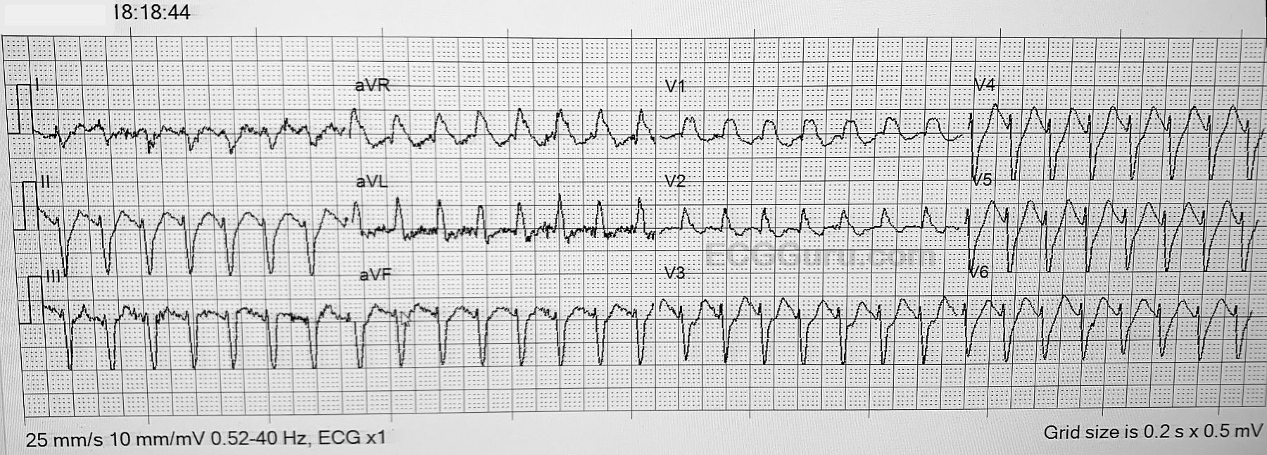
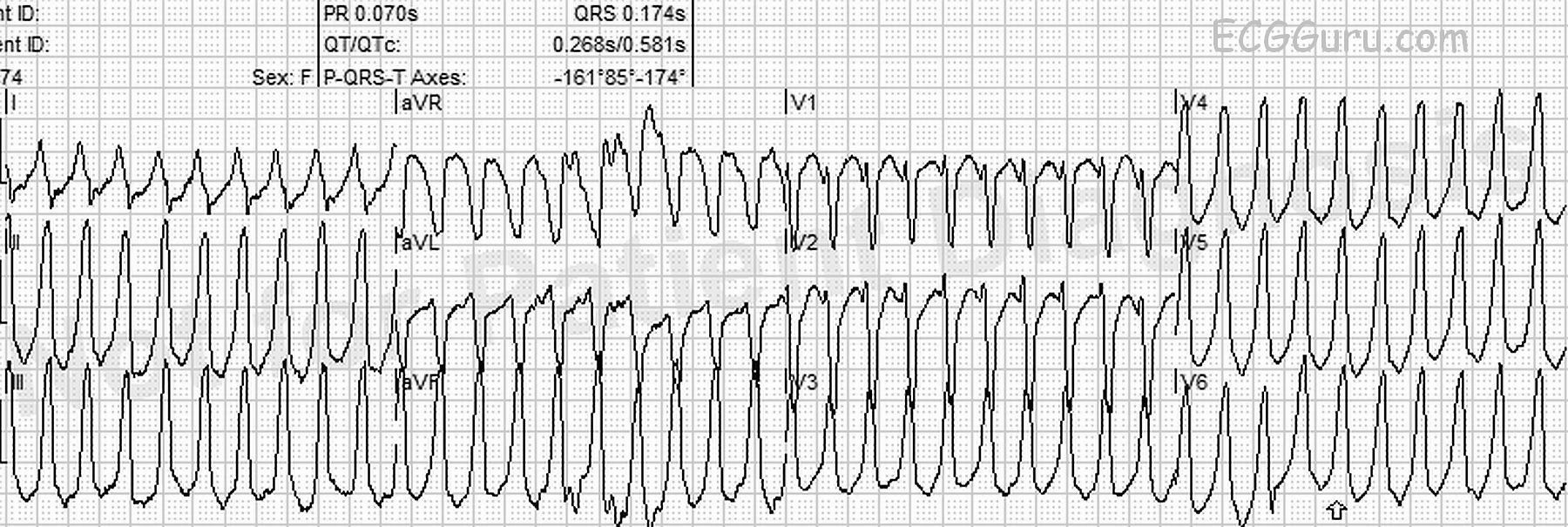
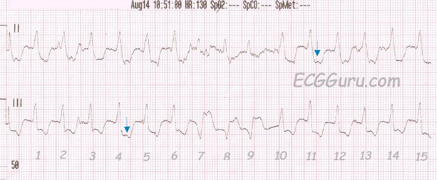
In the 12-Lead ECG, we see wide QRS complexes that are regular at a rate of 131 / minute. There are no obvious P waves before the QRS complexes, and no obvious distortion of the T waves, which would suggest a "hidden" P wave.  Unfortunately, there is significant artifact, which makes searching for P waves difficult.  The pattern overall suggests left bundle branch block, with the negative QRS in Lead V1 and positive QRS complexes in Leads I and V6.  However, one requirement for the diagnosis of LBBB is a supraventricular rhythm, and P waves are the best indicator of that.  An irregularly-irregular rhythm, indicating atrial fib, would also have made LBBB more likely.  In typical LBBB, the frontal plane axis is usually left-normal or left.  In this ECG, Lead III is taller than Lead I, putting the axis within normal range, but slightly rightward.

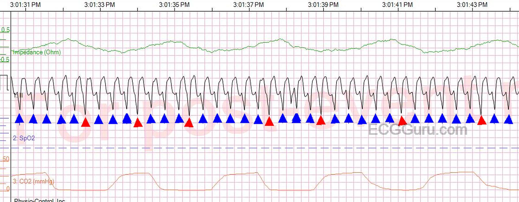
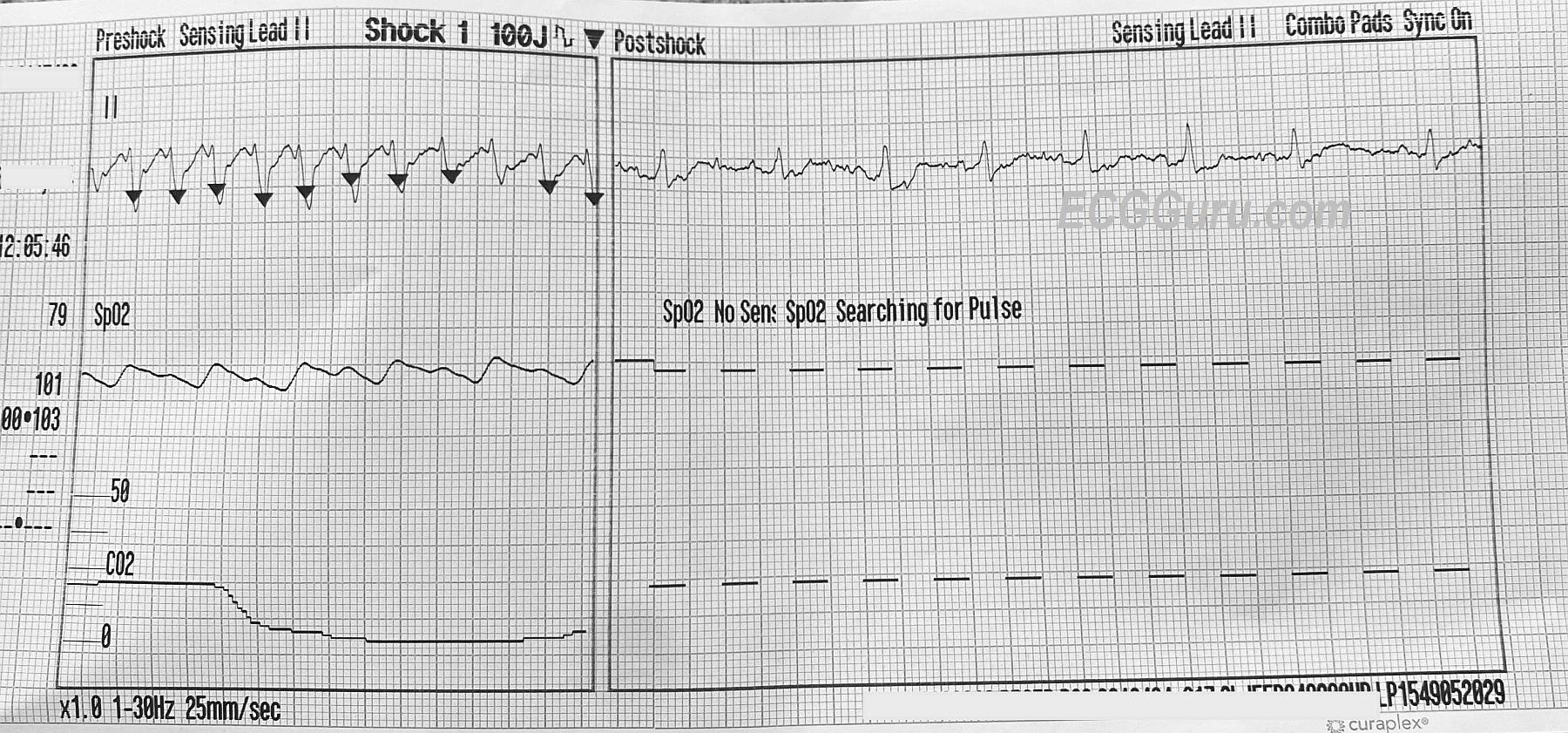
The rhythm strip uncovers something else.  Possible P waves are seen in some of the ST segments (arrows).  Are these dissociated?  Do they represent a first-degree AV block?  Are they actually artifact?  If this is a supraventricular rhythm, there is LBBB.   Then, notice beats #7,8,9.  If this rhythm is supraventricular (with LBBB), those must be a salvo of V Tach.  But, one of the possible P waves occurs in front of beat #7.

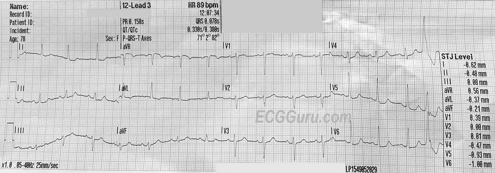
Another possibility is Right Ventricular Outflow Tract Tachycardia.  RVOT is a type of V Tach that typically has a LBBB pattern, with a slightly rightward axis.  If this is the case, beats #7,8,9 are probably "capture" beats or "fusion" beats.  Capture and fusion beats "prove" that the underlying tachycardia is ventricular, since, by definition, capture and fusion represent a return to supraventricular control of the rhythm.

Dawn's picture

Teaching Series: Atrial Fibrillation With Left Bundle Branch Block

This is a good example of atrial fibrillation with left bundle branch block.  You get two ECGs with this one, because the patient presented to EMS with a fast heart rate, and the rate was slowed with the drug diltiazem.  We do not have any other patient information, unfortunately.

In the first strip, we see a wide-complex tachycardia.  In an emergency situation, with an unstable patient, this rhythm could safely be treated using an emergency ventricular tachycardia (VT) protocol.  In fact, all WCTs should be considered to be VT until proven otherwise.  In most emergency settings, the unstable VT patient would be electrically cardioverted, which will often convert atrial fibrillation as well.  The stable patient with this rhythm would be treated with an antiarrhythmic drug, such as amiodarone.  This may convert or slow down atrial fib.  So, in the initial stages of treatment, differentiating between VT and A Fib is not the first priority.  Assessing the patient's hemodynamic stability and addressing the rate if necessary are the priorities.  

So, how do we know this is NOT VT?  It can be difficult, but in this case, the rhythm, even though fast, is very irregular.   VT is not always perfectly regular, but this irregularly-irregular rhythm points to atrial fibrillation. Also, the pattern of the QRS morphology fits with LBBB.  The criteria for LBBB are:  1) supraventricular rhythm, 2) wide QRS, 3) negative QRS in V1 with positive QRS in Leads I and V6.  If we assume the rhythm is atrial fibrillation, we meet the first criteria.  The other two are self-evident.

After the medication is administered, 10 minutes later, we see the rate slow down.  There is no change in the irregularly-irregular rhythm, and the LBBB pattern remains.  All that has changed is the rate and, hopefully, the patient's symptoms.  This confirms that the original rhythm was not VT.  

Remember, atrial fib lowers cardiac output because there is no P wave - no "atrial kick".  Also, the fast rate associated with new-onset atrial fib often compromises ventricular filling and cardiac output.   LBBB also has a deleterious effect on cardiac output.  Wide QRS complexes indicate that the ventricles are not contracting efficiently and synchronously.  The left ventricle is depolarizing by way of a slow wave of depolarization, rather than all the cells getting the message to depolarize at the same time.  Having these two conditions at the same time can have a very negative effect on cardiac output, leading to CHF.  The first step in treatment often involves simply slowing the rate to normal, which allows for better ventricular filling and decreases the workload on the heart.  Then, the fibrillation and bundle branch block can be addressed.

Dawn's picture

Wide Complex Tachycardia

This ECG shows a wide-complex tachycardia with a rate of 137/minute.  No patient information is available other than what is on the ECG.  Here, we will comment for the BASIC LEVEL learner, and allow the ECG Gurus out there to add INTERMEDIATE and ADVANCED level comments.

Beginners and emergency workers should ALWAYS be cautioned to treat all wide-complex tachycardias as VENTRICULAR TACHYCARDIA until proven to be something else.  V Tach is a life-threatening dysrhythmia, and there should be no delay in treatment.  That being said, not all WCTs are V Tach.  The most common "mimic" of V Tach is left bundle branch block.  LBBB can appear along with any supraventricular rhythm, including sinus rhythms, atrial tachycardias, atrial fibrillation, and junctional rhythms.  The diagnostic criteria for LBBB are:  wide QRS (.12 seconds or more), supraventricular rhythm, negative QRS in V1 and positive QRS in V6 and Lead I.  This ECG meets the criteria for LBBB, except that the supraventricular rhythm is hard to prove.  On one hand, P waves are not seen - or at least not easily.  On the other hand, the ECG machine has recorded a PR interval and a P wave axis.  What do you think?

Some of the criteria that would favor the diagnosis of V Tach are not present here: precordial concordance (all QRS complexes in V1 through V6 pointing in same direction), V6 with a negative QRS, AV dissociation.  An extreme axis, especially extreme right, would favor V Tach.  This ECG has an abnormal left axis, which is possible in V Tach AND in LBBB.   For a more thorough discussion by ECG Guru Jason Roediger of the criteria for differentiating V Tach from LBBB and other aberrancy, go to this LINK.

In an emergency setting, WTCs are most often V Tach.  However, should the rhythm turn out to be SVT with aberrant conduction, the usual V Tach treatment protocols would do no harm.  For instance, if the patient is deemed to be "unstable", electric cardioversion is recommended for both V Tach and SVT.  If the patient is stable, the emergency drugs usually recommended are generally safe for both rhythms.

Care should be taken to differentiate SINUS tachycardia from SVT or V Tach.  Sinus tachycardia usually has an obvious physiologic cause (hypoxia, hypovolemia, fear, pain, fever, etc.).  Sinus tachycardia also will be variable in rate, slowing as the cause is alleviated, or speeding if the problem causing the tachycardia becomes worse.  It may require  a long period of observation to determine that the rate is gradually slowing or speeding up.  One would hope to find P waves at some point in sinus tachycardia, and a 12-lead ECG is a great help.

WE LOOK FORWARD TO COMMENTS FROM OUR MEMBERS.

Dawn's picture

Wide Complex Tachycardia: Left Bundle Branch Block With Subsequent Rhythm Strip

 This wide complex tachycardia has an initial rate of 129/min. It is difficult to discern if P waves are present, although the ECG machine does give us a PR interval. The ECG meets most of the criteria for left bundle branch block: wide QRS, negative QRS in V1, positive QRS in Lead I and V6.The axis is leftward, which is common in LBBB.  However, it is difficult to say for certain that this is a supraventricular rhythm. Later, however, the patient's rate slowed (see top strip), revealing P waves. When the rate slowed, the left bundle branch block pattern remained. That helps confirm the original interpretation of left bundle branch block. Interestingly, the BBB is not rate-related, in that it is still present at the slower rate.

Of course, it would be helpful to have a complete 12-Lead ECG after the rate slowed, and it would also be good to see the onset and/or offset of the fast rhythm, which would help to determine if this is a sinus tachycardia or a paroxysmal supraventricular tachycardia (PSVT).  We do not have clinical data to help us determine if a sinus tachycardia with a rate of around 130 / min. would be appropriate or expected in this patient.

Click on image, or, for best image quality, right click and SAVE image.


Pages

All our content is FREE & COPYRIGHT FREE for non-commercial use

Please be courteous and leave any watermark or author attribution on content you reproduce.