Displaying 1 - 6 of 6
Dawn's picture

Severe Triple Vessel Disease

The Patient:   This ECG is from a 63-year-old man who complained of epigastric pain for three hours. The pain was sudden in onset, burning in nature, and accompanied by nausea and palpitations.  The patient is a heavy smoker, diabetic and hypertensive with a long history of non-compliance to his medications. 

He was given crushed aspirin, loaded with clopidogrel and heparin, given high-intensity statins, and rushed to the cath lab. 

The ECG:  The rhythm is normal sinus, a bit fast at 90 bpm.  The intervals, frontal plane axis, and R wave progression are normal.  This ECG shows a very dreaded pattern:  ST segment elevation in aVR and V1 with widespread ST depression, seen here in all other leads.  This is an ECG sign of GLOBAL ISCHEMIA.  There are several possible causes, all bad.  The most common causes of this pattern are:

·        Severe triple vessel disease, with significantly decreased flow in the left anterior descending, right, and circumflex arteries.

Dawn's picture

Anterior-lateral M.I. With Wide QRS

The Patient:  An elderly man presents with chest pain, pallor, diaphoresis and weakness.

The ECG:     The rhythm is normal sinus at a rate of about 76 bpm with normal intervals. The QRS complexes are wide at about .14 seconds (140 ms).  There is ST segment elevation in all precordial leads, except for possibly V6.  The shape of the ST segments in the anterior wall range from coved upward in a “frowning” shape (V1) to very straight (V5 and V6).  There is also ST elevation in aVL with ST straightening in Lead I.  There is ST depression in the inferior leads, II, III, and aVF.  Lead II is equally biphasic while I and aVL are positive, indicating an axis that is shifted slightly to the left.  With his symptoms and this alarming ECG, he was sent promptly to the cath lab.

Interpretation:  The rather obvious ST-elevation M.I. is extensive, covering the entire anterior wall, and extending into the high and low lateral walls . This was confirmed in the cath lab, as the patient had an occlusion of the left anterior descending artery near the bifurcation of the circumflex.  The wide QRS meets the criteria for left bundle branch block (wide QRS, negative QRS in V1 and positive QRS in V6 and Lead I).  However, it doesn’t have the “look” of LBBB with the low-voltage seen in the anterior wall. After the offending artery was opened and stented, the wide complex became narrow and was considered to be an interventricular conduction delay that was due to the ischemia.  The ST depression in the inferior wall is most likely reciprocal.

Dawn's picture

Ask The Expert

Today’s expert is Dr. Jerry W. Jones, MD, FACEP, FAAEM

Jerry W. Jones, MD FACEP FAAEM is a diplomate of the American Board of Emergency Medicine who has practiced internal medicine and emergency medicine for 35 years. Dr. Jerry JonesDr. Jones has been on the teaching faculties of the University of Oklahoma and The University of Texas Medical Branch in Galveston. He is a published author who has also been featured in the New York Times and the Annals of Emergency Medicine for his work in the developing field of telemedicine. He is also a Fellow of the American College of Emergency Physicians and a Fellow of the American Academy of Emergency Medicine and, in addition, a member of the European Society of Emergency Medicine. 

 Dr. Jones is the CEO of Medicus of Houston and the principal instructor for the Advanced ECG Interpretation Boot Camp and the Advanced Dysrhythmia Boot Camp. 

QUESTION:  How can I explain to students that injury from an M.I. “localizes” on the ECG, but subendocardial ischemia/injury does not?

ANSWER:                 Allegory of Subendocardial Ischemia

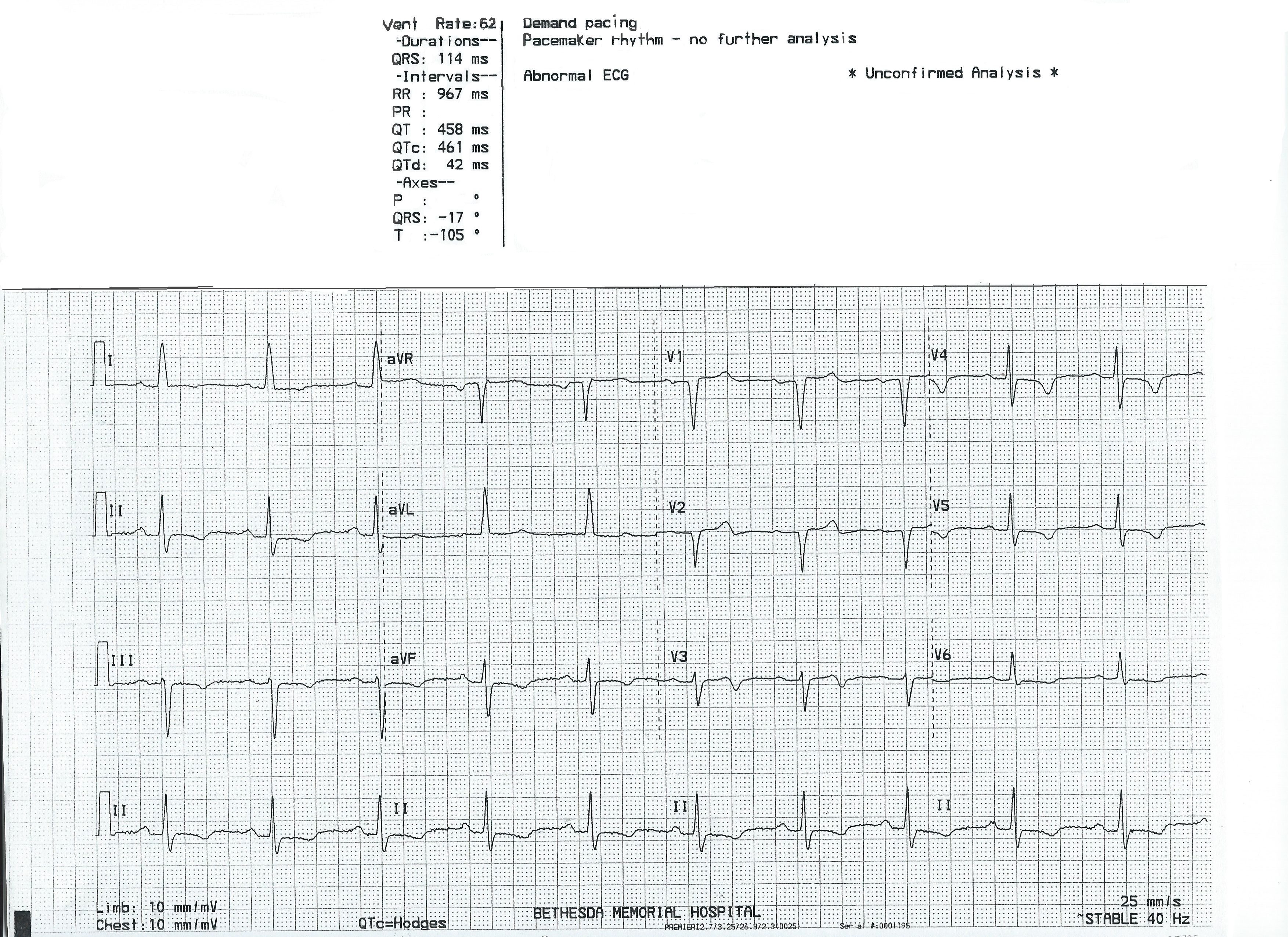
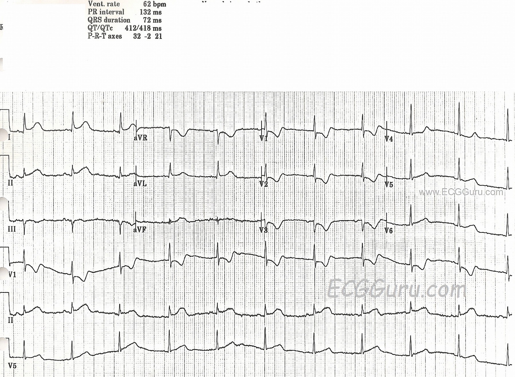
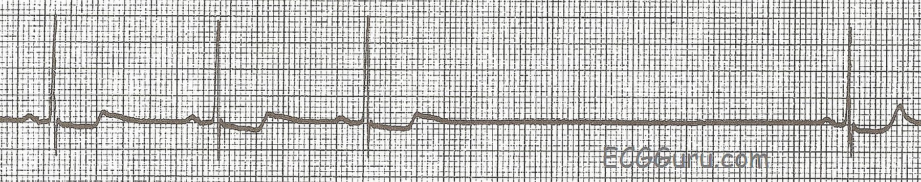
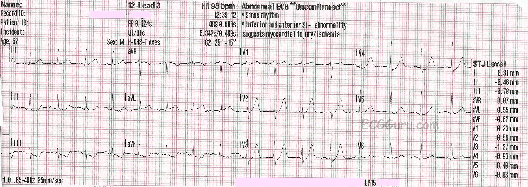
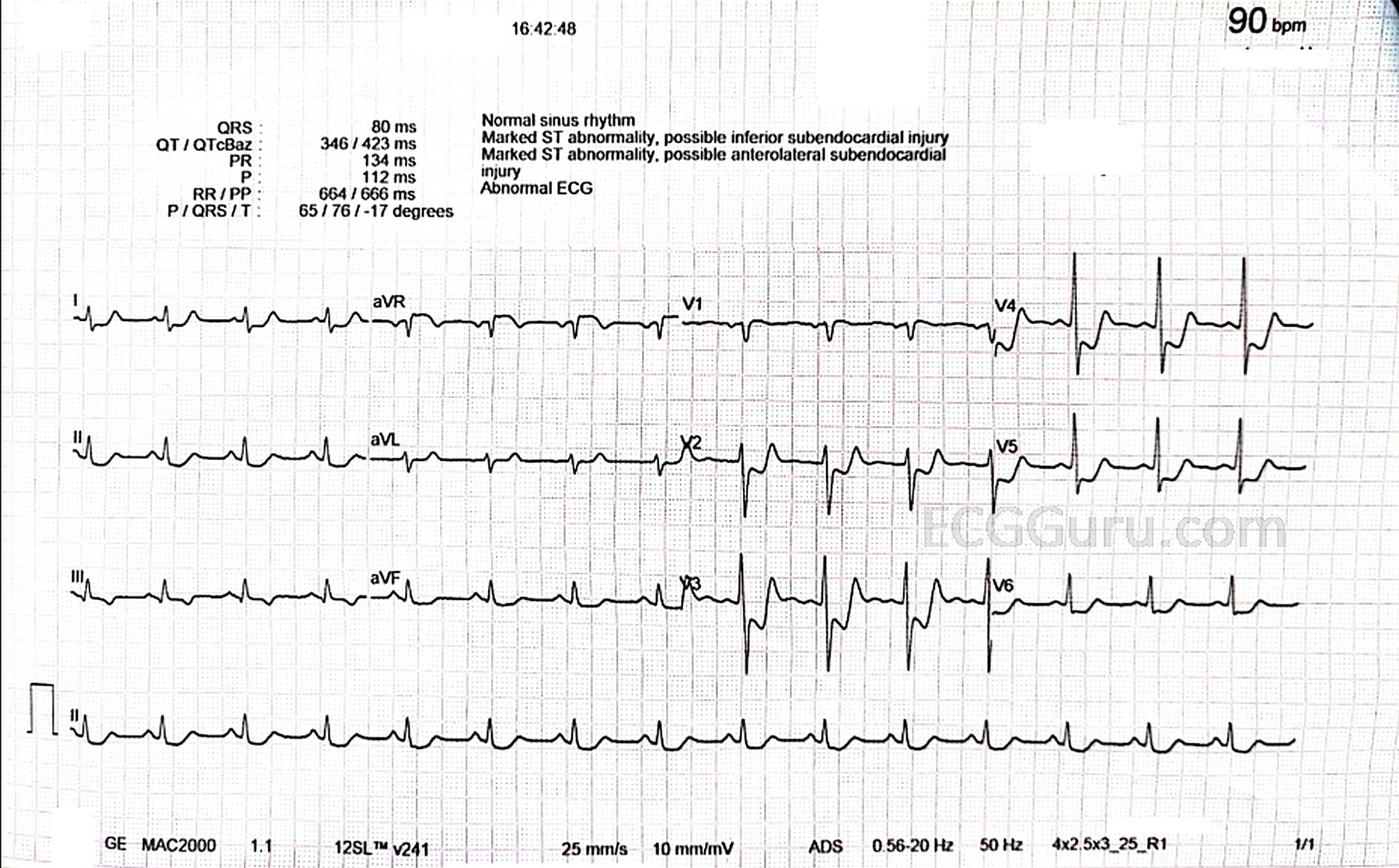
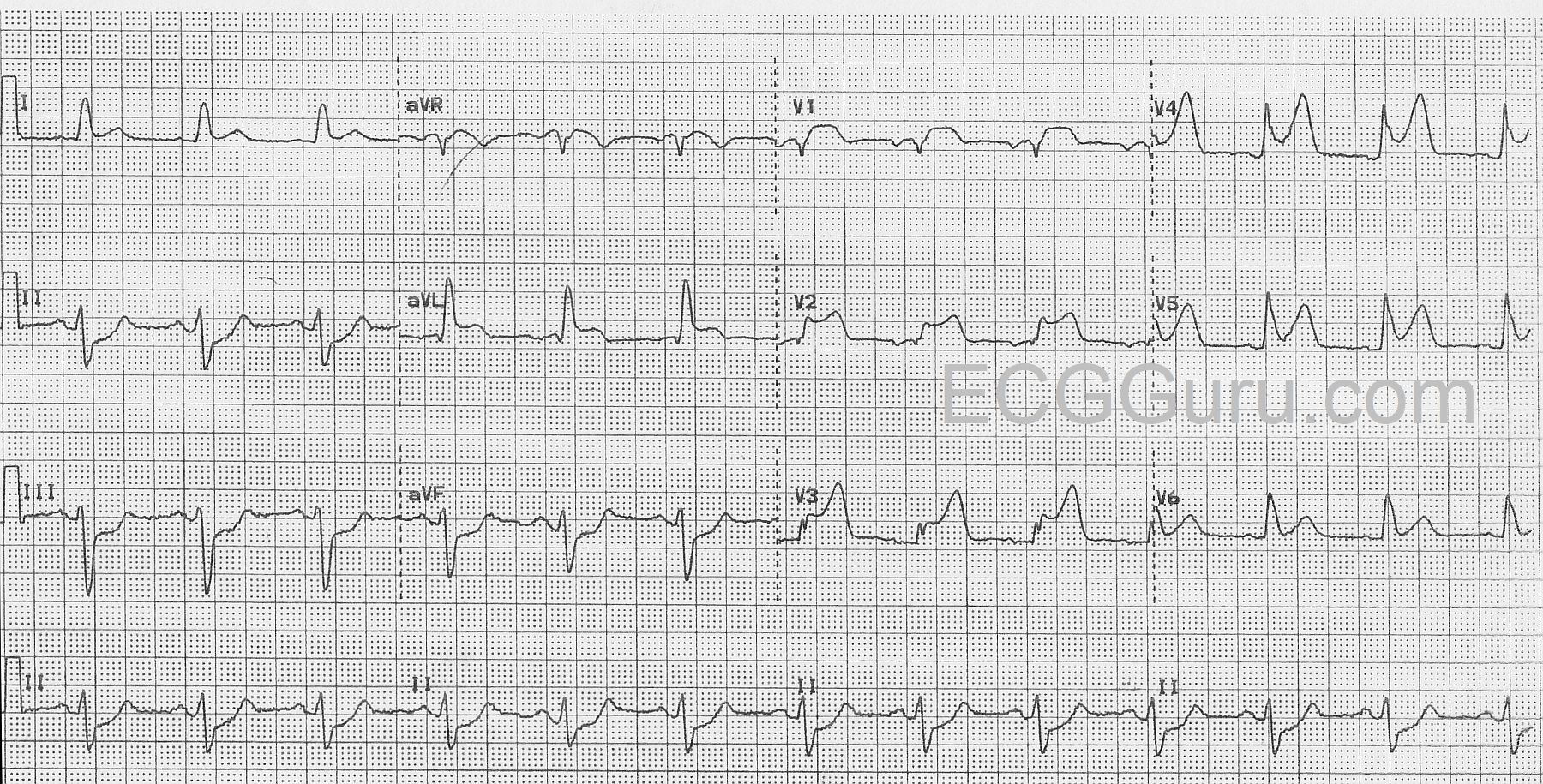
 For many years we have misunderstood the concept of subendocardial ischemia as it manifests on the 12-lead ECG.  Previously, if one saw ST depression in leads II, III, and aVF, it would be labelled "inferior subendocardial ischemia" and, if the patient were momentarily having little or no chest pain, the patiet would be sent home.  The same thing happened with ST depression in leads V4 - V6; "anterolateral subendocardial ischemia," probably chronic and again, the patient may be sent home.  And of course, ST depression in leads V1-V4: "anteroseptal subendocardial ischemia" and often the patient was sent home.

Then a number of years ago, some disturbing information began to surface in various medical journals around the globe.  Sometimes ST depression that was limited to just leads II, III, and aVF, for example, did not reveal any actual subendocardial ischemia in the inferior wall of the left ventricle.  In some cases, subendocardial ischemia was indeed present but very little involved the inferior wall and most of the ischemia was elsewhere; but... the only ST depression present was in the inferior leads.  Some articles began mentioning the same findings regarding ST depression in other leads as well.

What we have learned is that when ST depression indicates subendocardial ischemia, IT DOES NOT LOCALIZE!  Just because there is ST depression in leads II, III, and aVF does NOT necessarily mean that the ischemia is located in the inferior wall of the left ventricle.  It MAY be there, or there may be SOME ischemia there but most of it elsewhere, or there may be NO ISCHEMIA AT ALL there.

Some people still have difficulty conceptualizing this, so I developed an allegory of subendocardial ischemia using the concept of a vacant house at night.


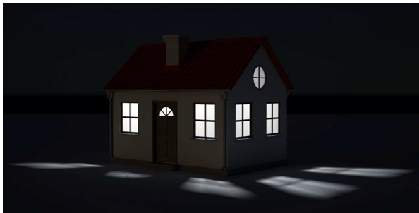
Imagine you have gone out for a walk one pleasant evening in your neighborhood.  As you stroll down the street, you come upon a vacant house.  You know it is vacant because the family that lived there moved out recently.  However, you can see light coming from some of the windows of the house. You wonder what's going on, so you walk up to the house and look through a window into the living room.  The room is illuminated but you don't actually see a light on there.  You move around to another window and look into the dining room.  Again, there is enough light for you to see everything in the room but you don't actually see any light fixture that is on. Finally, you move around to the window that looks into the kitchen.  It's illuminated as well and you can see everything but, once again, the source of the light is not there.  Is the light in a room that you cannot see or is it perhaps a closet light that has been left on somewhere in the house?

That - in essence - is subendocardial ischemia.  Just because you see ischemia through the "windows" of leads II, III or aVF or the "windows" of leads V4 - V6 doesn't mean that the "source of the ischemia is in those "rooms."

Subendocardial ischemia manifested by ST depression does NOT localize reliably.  So how should you report such ischemia?  This is what I would say if I saw (for instance) ST depression in leads V4 - V6:  "There is subendocardial ischemia present indicated by ST depression in leads V4 - V6."  I would NOT call it "anterolateral ischemia."

Actually, this information has been available for a number of years.  So, if you are reading textbooks, journal articles or posts on websites that still refer to "inferior ischemia", "high lateral ischemia", "anteriorlateral ischemia" etc., then you are reading information that is OUTDATED. If it was recently written, then you are reading information from someone who is NOT staying current with advances in electrocardiography.


Dawn's picture

Incorrect Machine Interpretation

This ECG is presented as an example of INCORRECT MACHINE INTERPRETATION.  While there are many abnormalities in this ECG, it does not represent a paced rhythm. While there are exceptions, most paced rhythms represent either AV sequential pacing, right ventricular pacing, or bi-ventricular pacing.

RECOGNITION OF A PACED RHYTHM

Recognizing a paced rhythm can be difficult in some cases. Because pacemakers now have so many programmable features, there is a wide variety of ECG changes associated with them.  Pacer “spikes” can be difficult to see in all leads.  Finding evidence of the device on the patient’s chest or via patient history is a big help in reminding us to scrutinize the ECG for paced rhythm.

An AV sequential pacemaker or a right ventricular pacemaker will pace the ventricles via the right ventricle.  This produces a WIDE QRS and a leftward axis, often causing Leads II, III, and aVF to be negative and aVL and aVR to be positive.  Along with the wide QRS, we will see DISCORDANT ST CHANGES.  That is, there will be ST depression and T wave inversion in leads with positive QRS complexes and ST elevation and upright T waves in leads with negative QRS complexes.

Bi-ventricular pacing can be a little more complicated to recognize, as the QRS can be narrow, with signs of fusion between the wave produced by the LV electrode and the RV electrode.

The frontal plane axis is usually far right – aVR will be positive.  Lead I will be negative.

The machine is wrong:  there is no indication of a pacemaker, and P waves are present, even though they are not noted in the "PR Interval" or "P Axis".

SO, THIS IS NOT A PACED RHYTHM – WHAT IS IT?

Dawn's picture

Circumflex Occlusion with Posterior-lateral M.I.

This ECG was obtained from a woman with chest pain who was taken to the cath lab and found to have a 100% occlusion of her circumflex artery.  

There are obvious ST segment elevations in Leads I and aVL, as well as in Lead II.  Lead II is the most leftward of the inferior wall leads, and I and aVL reflect the high lateral wall. She also has ST depressions in V1 through V3.  If you look closely at the R wave progression in the anterior leads, you will readily note that it appears that V1 and V3 wires have been reversed.  That being said, the "real" V2 and V3 have taller-than-normal R waves.  The tall R waves and ST depression are signs of "posterior wall M.I."  Recently, the actual definitions of the "lateral" wall and "posterior" wall have come into question.  However, the important thing clinically, is that this patient IS experiencing an ST elevation M.I. (STEMI), which was confirmed in the cath lab.  The locations of the ST changes were consistent with the 100% occlusion of her circumflex artery.

For our more advanced readers (and our "Gurus"), there is an interesting rhythm.  The P wave morphology changes frequently, even though the rhythm remains regular.  The rate, at 62 BPM, was adequate, and the patient did not suffer any consequential dysrhythmias during her procedure.  We don't have long-term followup information on her.


Dawn's picture

ECG Basics: Sinus Pause / Sinus Arrest

This example of sinus arrest, also called sinus pause, shows a spontaneous return to sinus rhythm.  There are many mechanisms by which pauses can occur on the ECG. One concept for beginner students to grasp is that, if the pause contains the equivalent of regular R-to-R intervals, and the first complex after the pause is "on time",  we can expect that the sinus node kept firing, but did not penetrate the atria (exit block). If the pause is irregular in length, with the first beat after the pause seeming to come in randomly, we can call this sinus arrest or pause, understanding that there are many different mechanisms that can be at work here. Because what little we can see of the underlying sinus rhythm is irregular, or speeding up, we cannot discern absolutely that this is sinus arrest. 

The bottom line for the patient, and for any level practitioner, is, "how is the patient tolerating this pause, and what does it mean to the patient's overall prognosis?  In the short term, the patient may require emergency pacing while the cause of the dysfunction is investigated. If pauses are long enough to lower cardiac output, they can cause fainting. More than a few automobile accidents have happened as a result of this type of dysrhythmia. 

For more advanced students, this short rhythm strip (Lead II) reveals sloping ST depression, and indicates the urgent need for a 12-lead ECG and other tests. 


All our content is FREE & COPYRIGHT FREE for non-commercial use

Please be courteous and leave any watermark or author attribution on content you reproduce.