
Submitted by Dr A Röschl on Fri, 07/14/2023 - 04:07
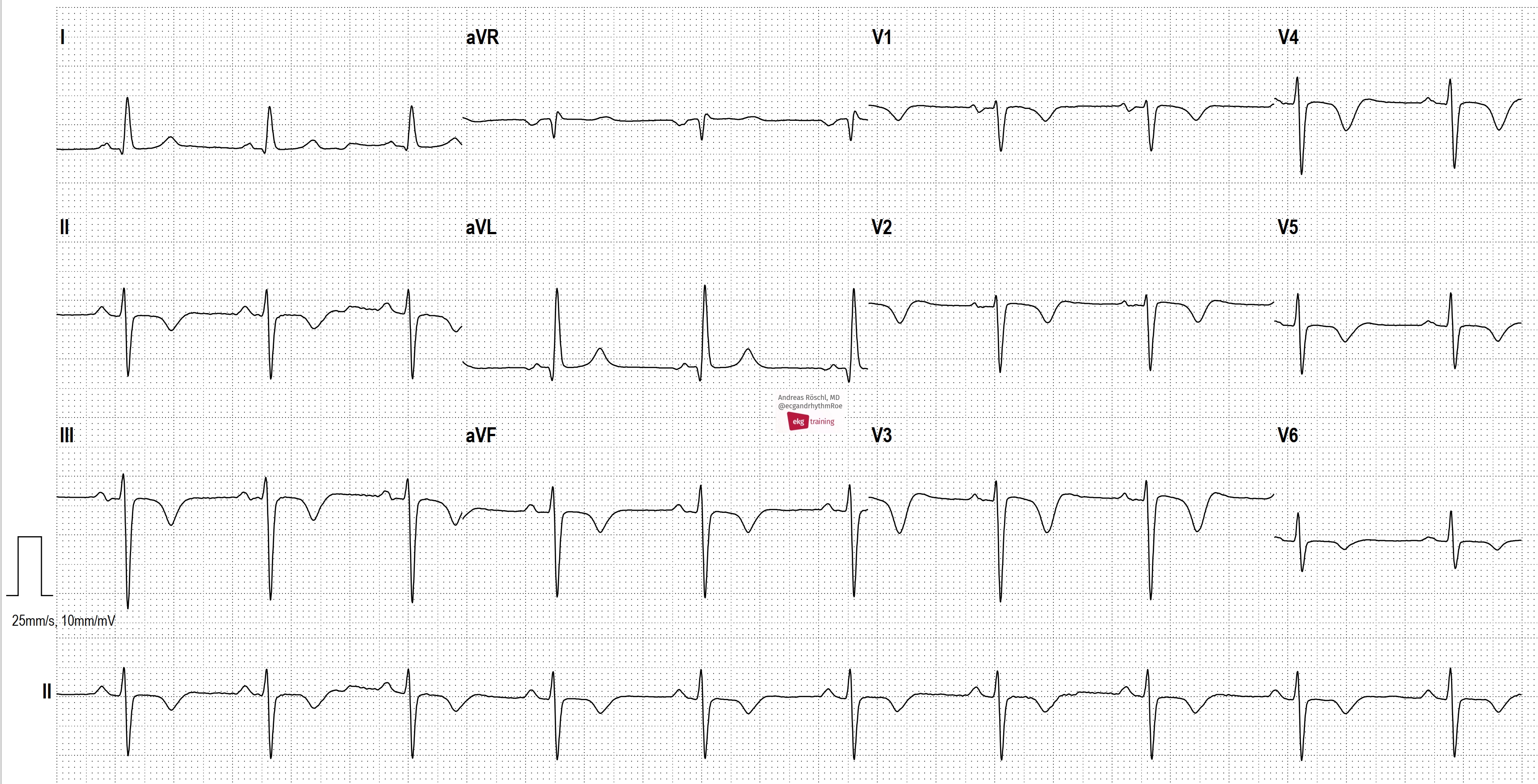
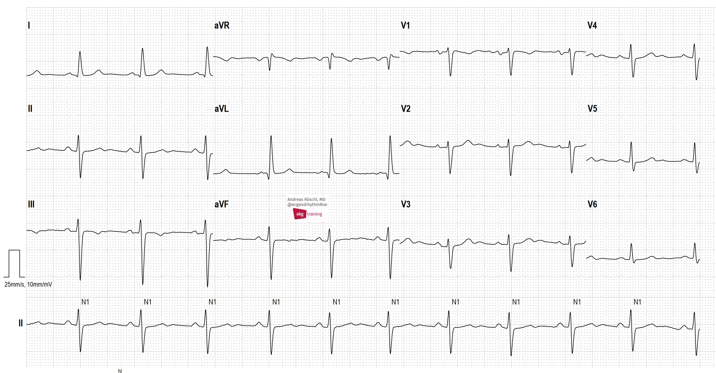
ECG 1 is from a 57-year-old male with no prior cardiac disease. He reports acute shortness of breath for 2 days. We see a sinus rhythm with left anterior fascicular block (LAFB) and conspicuous T-wave inversions in the inferior leads and in V1-V6. These are typical ECG changes that may indicate a pulmonary embolism. ECG 2 was taken from the same patient 1 year earlier. The patient has an acute pulmonary embolism. Sinus tachycardia may be present in acute pulmonary embolism. However, as in this example, the heart rate can also be completely normal.
Rate this content:
-

- Dr A Röschl's blog
- Log in or register to post comments
All our content is FREE & COPYRIGHT FREE for non-commercial use
Please be courteous and leave any watermark or author attribution on content you reproduce.
Comments
Good Reminder
Dawn Altman, Admin