Submitted by Dr A Röschl on Fri, 01/03/2025 - 02:39
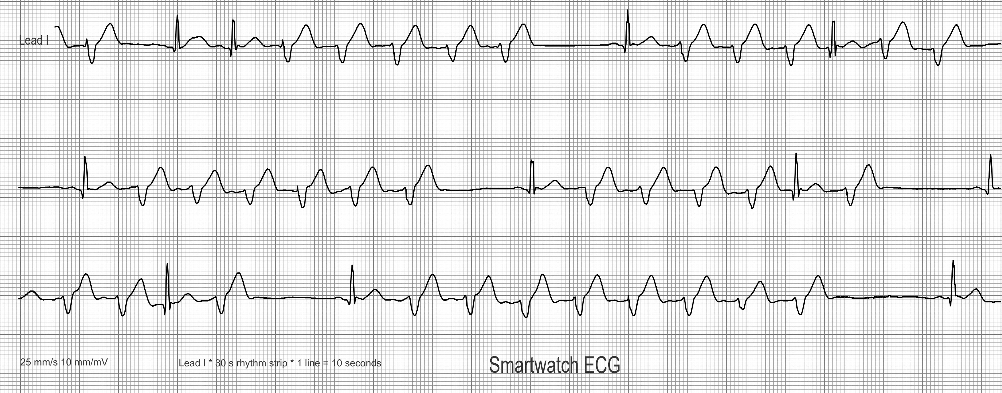
Here we see a 30 s long strip of a 1-lead ECG rhythm strip, recorded with a modern Smartwatch (Apple watch). The ECG has been graphically processed for better visualization. The paper speed is 25 mm/s as usual. The lead shown corresponds to ECG lead I of the limb leads.
Submitted by Dr A Röschl on Sun, 05/28/2023 - 02:21
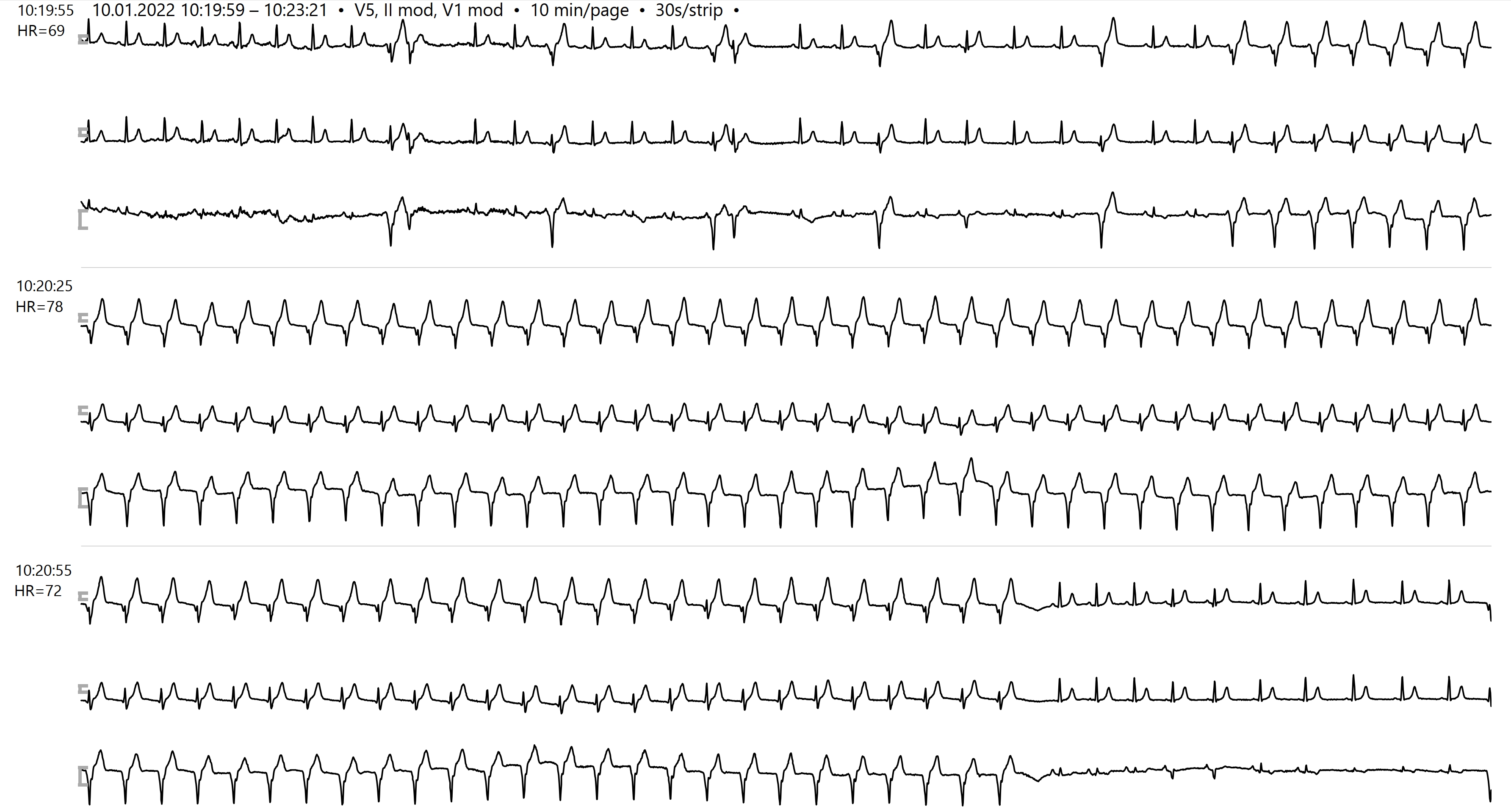
This Holter-ECG comes from a 70-year-old physician with coronary artery disease. He is twelve months after PCI (RCA, RCX). Currently no symptoms (no dyspnea, no chestpain, no palpitations). You can see that an overview/section from a Holter- EKG, 3 channels are shown, strip width is 30 seconds. First you can see a sinus rhythm with frequent PVCs, then there is a transition to an accelerated idioventricular rhythm (AIVR) that lasts almost a minute in total.
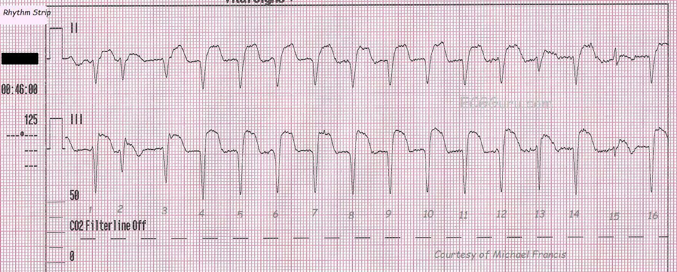
This rhythm strip is taken from a 67 year old man with chest pain who had ECG signs of inferior-posterior wall M.I. upon the arrival of paramedics. He had a recent (5 day) history of cardiac cath and stent placement. During his treatment, his condition and ECG signs improved somewhat, and it was felt that he spontaneously reperfused during transport. Often, during reperfusion of the heart after a total or near-total occlusion, dysrhythmias will appear. They are usually transient. This strip shows accelerated idioventricular rhythm. The criteria are: wide complex rhythm with no P waves associated, rate between 40 and 100 bpm. The rate can go higher. Some people would prefer the term V Tach when the rate is over 100. This rhythm, however, is not usually as sinister as V Tach.Sometimes, it can be difficult to differentiate AIVR from sinus rhythm with hidden P waves and a conduction defect such as bundle branch block, which widens the QRS. Artifact such as we see here can obscure P waves, as can a very fast rate. The real clue to this being AIVR is the "capture" beat - number 15 - at the end. This is a sinus beat, proving that there is a separate underlying sinus rhythm. Beat number 2 is a fusion beat - the ventricular beat coming from below has "collided" with the supraventricular depolarization wave from above (in this case a PAC). The resulting QRS has characteristics of both beats. This AIVR has taken over the heart, as the FASTEST PACEMAKER CONTROLS THE HEART. To see a series of 12-lead ECGs from this patient, go to http://ecgguru.com/ecg/teaching-series-112213-inferior-posterior-wall-mi...