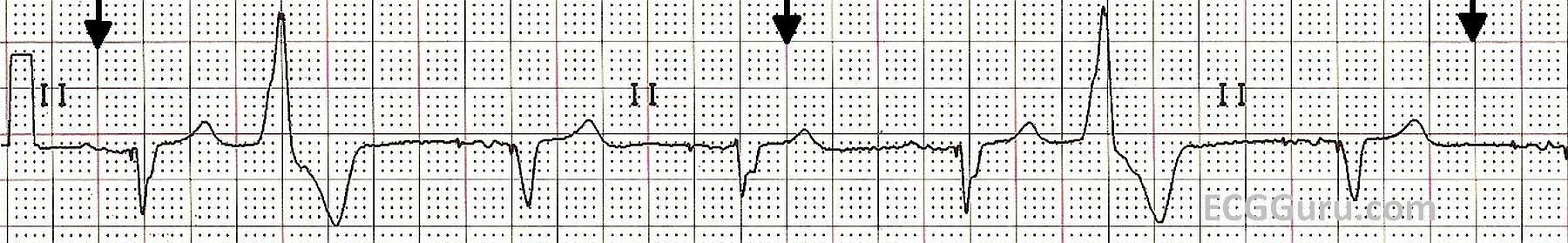
This is a Lead II rhythm strip from a patient with a biventricular pacemaker. The second and sixth beats are PVCs. In this lead, the pacemaker spikes are very difficult to see, but they are present. The pacemaker is operating in a "demand" mode for pacing the atria. Some of the P waves appear to be the patient's own, and some appear to be caused by the pacing stimulus. For example, the first beat appears to have no pacer spike before the P wave, and the second beat does have one (albeit tiny). The morphology of the P waves appears to change, also.
This pacemaker has been programmed to cause a QRS complex after every P, whether the P wave was made by the patient or by the pacemaker. The patient originally had a left bundle branch block, making his QRS complexes very wide, and lowering his cardiac output. The biventricular pacemaker paces both ventricles, synchronizing their depolarization and narrowing the QRS. This improves cardiac output. The physician has programmed this pacemaker to pace the ventricles after every P wave, whether native or paced. The paced QRS happens slightly before the native (wide) QRS would have, giving the patient the benefit of narrow QRS complexes. Biventricular pacemakers have been shown to improve cardiac output in patients with wide complexes.