Displaying 1 - 3 of 3
Dawn's picture

Left Bundle Branch Block

This is a good example of sinus rhythm with left bundle branch block.  There is some irregularity due to a PAC at the beginning.  The QRS is wide at 144 ms (.14 seconds).  There is also first-degree AV block, with a prolonged PR interval of 228 ms.  The criteria for diagnosis of left BBB are:  wide QRS, supraventricular rhythm, and a negatively-deflected QRS in V1 with a positive QRS in Leads I and V6.  

Left bundle branch block can be associated with many forms of heart disease, including CHF.  It can be permanent, transient, intermittent, or rate-related.  The wide QRS of LBBB significantly decreases cardiac output, causing poor perfusion symptoms in some people.

This ECG is a good one for your students who are just transitioning from reading rhythm strips to reading 12-lead ECGs.  It shows the value of multi-lead assessment of rhythms. You will notice that P waves are difficult to see in some leads.  Armed with the knowledge that the four channels on this ECG are run simultaneously, you can show the students how finding P waves in one lead will allow you to find them in the leads that are above and below that lead. 

Similarly, it can be difficult to see the QRS width in some leads.  The leads in the same vertical column can help you see the QRS's true width, even if part of the QRS is "flat" in the isoelectric baseline.

Dawn's picture

Teaching Tip: 12 Leads are Better Than One (Or Three)

Years ago, I was tasked with introducing 12-lead ECG interpretation to firefighter/paramedics who had been using ECG for rhythm monitoring for years.  Some were eager to add to their skills, others - not so much.  The feeling was, we have been doing just fine as we are.  When finally convinced that they could interpret STEMI with a 12-lead, many were content to use the 12-lead ECG only for that.  

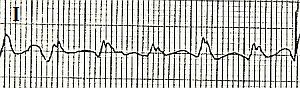
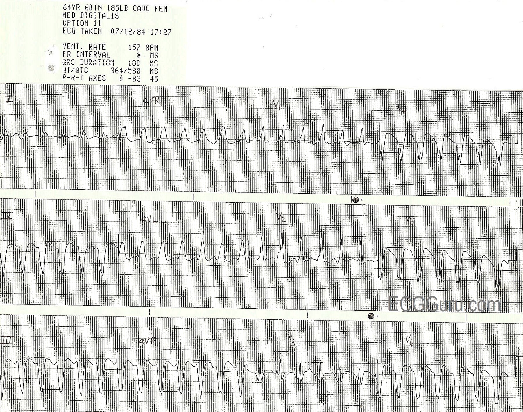
To illustrate to students the great value of multi-lead assessment, I devised a little "quiz".  I showed the students ten to twelve short rhythm strips, like you see here.  All were cropped from 12-lead ECGs.  I asked my class to interpret the strips as they would if they were taking an ACLS class.  Usually, all did fine, or so they thought.  When shown the 12-lead ECGs the strips were taken from, EVERY student changed his or her mind on EVERY ECG.  The lesson is:  sometimes what we are looking for shows up in some leads and not others.  You can find this illustrated hundreds of times just in the ECG archives on this site.  I will supply some ECGs here on this page over the next few weeks that you could use to show your own students the value of "multi-lead assessment".  

What started as a hard-sell turned out to be a fun exercise.

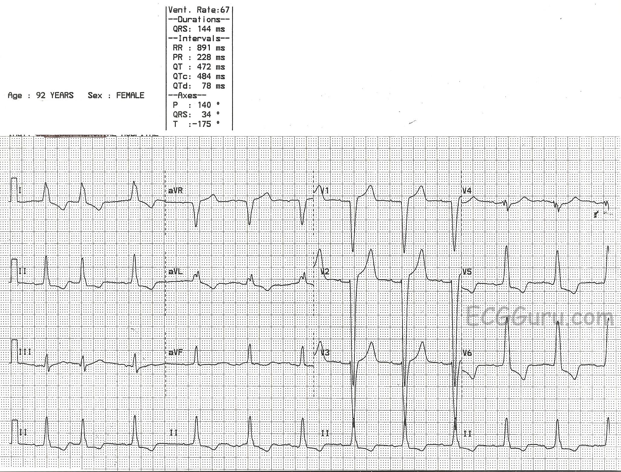
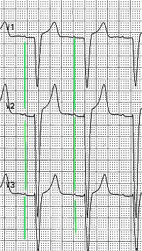
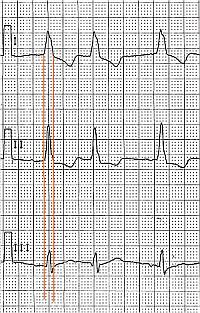
The ECG shown here is of a patient in V Tach.  There are several strong signs that this is V Tach, including the wide QRS complexes, lack of associated P waves, "backward" axis, also called extreme right axis deviation (Leads II, III, and aVF are all negative and aVR is positive), and V6 is negative.  For more review of the differential diagnosis of wide-complex tachycardias, go to our Ask the Expert answer from Jason Roediger.  This LINK willl take you to Dr. Grauer's informative webpage where he offers a step-by-step guide to differentiating the WCTs.

 The focus of THIS lesson is that, while the patient is in V Tach, and it is in every lead, the tell-tale signs are harder to see in some leads than others.  Remember to show your  Remember to share with your students that the channels of the ECG (in this case three) are run simultaneously, so that the same heartbeat is seen several times - once for each channel.

Dawn's picture

Left Bundle Branch Block

This ECG offers a good example of the left bundle branch block pattern.   * The QRS is wide at 144 ms.  * There is a supraventricular rhythm - in this case, normal sinus rhythm with beat number two a PAC, and a slightly prolonged PR interval.  * The QRS is negative in V1 and positive in V6 and also Lead I.  This satisfies the ECG criteria for left bundle branch block.

The main lesson this tracing offers for beginner or refresher students is the value of multi-lead assessment. Using only one or two leads, you may miss important information needed to correctly interpret the ECG.   In order to meet the LBBB criteria, we must show that there exists a supraventricular rhythm (not ventricular). One easy way to prove the rhythm is supraventricular is the presence of P waves.  In this ECG, P waves are very small, and are invisible in some leads, such as Lead I, aVL, Lead III, and aVF.  P waves can be seen well in Lead II and in the chest leads.  Some helpful hints, if viewing on a computer, enlarge the image to better see the P waves.  Look at the ECG machine's interpretation.  If a numerical PR interval is given, and a P wave axis, then the computer is finding P waves.   If you don't see them in one lead, try others.

For your more advanced students, ST elevation is common in wide-QRS rhythms, occuring in leads that have a negative QRS complex.  Conversely, ST depression will be seen in leads with wide, upright QRS complexes.  This makes the ECG with LBBB very confusing to read.  Evaluation of the ST segments should be deferred to experts.  Most EMS field protocols allow for a STEMI Alert to be called in LBBB only if the LBBB is known to be new-onset and the patient has obvious cardiac symptoms.  You can find many more examples of LBBB on this site, even LBBB with acute MI.   

All our content is FREE & COPYRIGHT FREE for non-commercial use

Please be courteous and leave any watermark or author attribution on content you reproduce.