Displaying 1 - 2 of 2
Dawn's picture

Large Anterior Wall M.I. and Effect of Lead Reversal

EDIT: Please refer to the comments below this text. The second ECG in this series shows unexpected QRS and ST-T morphology changes, which I tried to explain by way of the patient's long anterior descending coronary artery. However, Dave Richley, who is a very well-known cardiac physiologist and ECG Guru took the time to analyze these morphologies and realize they can be explained by an inadvertent ECG LEAD MISPLACEMENT. This patient does have a proximal lesion of the LAD, proven and repaired in the cath lab. But the inferior wall does not have the injury it appears to have in this second ECG. Thanks to Dave for reminding us to slow down and look closely when things don't look "right".

The Patient:   These two ECGs, taken 26 minutes apart, were obtained from a 50-year-old man who complained of sudden onset of chest pain.  He denied history of coronary artery disease. He was Covid-positive, and the rest of his medical history was unremarkable.

ECG No. 1:  This ECG was obtained by paramedics enroute to the hospital.  For your beginner-level students, it will be easy to demonstrate the large ST elevations in V3 through V6. The machine’s measurements at the bottom confirm that this ECG meets any field criteria for ST elevation M.I. “STEMI”.

But there is so much more to see! Taking a methodical approach, and starting with rate and rhythm, we see sinus bradycardia at 57 bpm. Intervals and frontal plane axis are within normal limits. R wave progression in the chest leads is stalled in V1- V3 due to loss of initial r waves (narrow QS). The transition to positive deflections in V4 – V6 is abrupt.  These q waves in the V1 and V2 appear narrow, but V3 appears to have a Q wave that is almost wide enough to be considered pathological.  Narrow Q waves may be a transient sign of injury, while wide ones (>40 ms) are an ECG sign of necrosis.

Dawn's picture

Acute Anterior-Lateral Wall M.I.

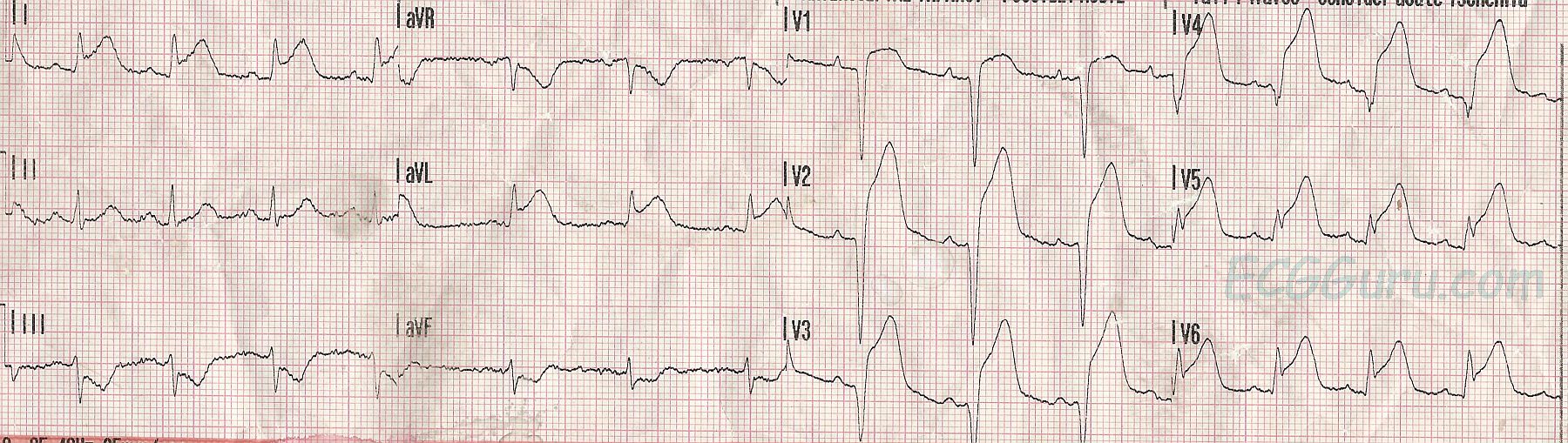
This week's ECG is from a 47-year-old man who experienced a sudden onset of chest pain while mowing his lawn.  He went on to suffer a cardiac arrest and was resuscitated.  We do not have long-term followup on his outcome.

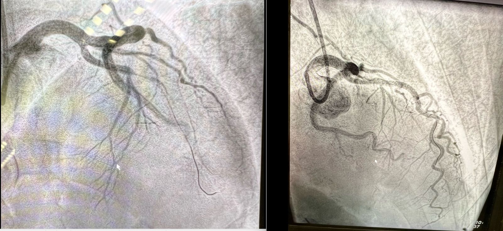
The experienced person will have no difficulty identifying a large acute antero-lateral wall M.I.  There are massive ST segment elevations in Leads V1 through V6, reflecting acute injury from the septal side of the anterior wall (patient's right) to the anterior-lateral wall (patient's left).  There are also ST elevations in Leads I and aVL, reflecting the high lateral wall.  This indicates, and was confirmed in the cath lab, that the lesion is proximal - at or above the bifurcation of the left anterior descending artery and the circumflex artery.  The ST depressions in the inferior wall leads (II, III, and aVF) likely represent reciprocal changes.  You will note that the ST depression in Lead III has a very similar shape to the ST elevation in Lead aVL.

More bad news for this patient is the presence of pathological Q waves in Leads V1 through V4, reflecting transmural death of the myocardial tissue.  This causes akinesis and poor left ventricular function.  In addition, it's not only muscle tissue that dies, but also electrical structures , such as bundle branches.   Papillary muscles can be infarcted, causing valve malfunction.  And remember, all patients who have ST elevation due to acute injury are vulnerable to ventricular tachycardia and ventricular fibrillation, due to re-entry mechanisms in injured tissue.   

This ECG will allow instructors to discuss with their students:

*  which leads reflect changes from which parts of the heart

*  what the ECG signs of acute M.I. are

*  the pathophysiology of pathological Q waves

*  the effect of damage to various parts of the heart on the patient's condition and symptoms

This "classic" M.I. pattern should be taught to all health care professionals who work in settings where ECG is used.

All our content is FREE & COPYRIGHT FREE for non-commercial use

Please be courteous and leave any watermark or author attribution on content you reproduce.