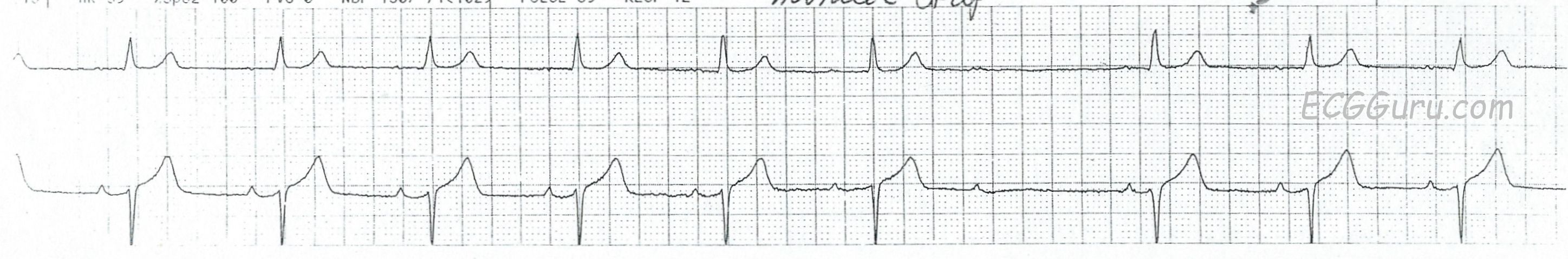
This two-lead rhythm strip shows a normal sinus rhythm at about 63 bpm. The P waves are regular. After the sixth P-QRS, there is a non-conducted P wave. The normal rhythm then resumes. The two most common reasons for a non-conducted P wave in the midst of a normal sinus rhythm are 1) non-conducted PAC, and 2) Wenckebach conduction. The first is easy to rule out. The non-conducted P wave is not premature, so it is not a PAC. The second one is a little harder when we only have a short strip to look at. We are conditioned to look for progressively-prolonging PR intervals until a QRS is "dropped". In this case, the progression is in very tiny increments that are hard to see unless you zoom in and measure. But they ARE progressively prolonging. An easy hack: measure the last PRI before the dropped beat and the first one after the pause. You will see that the cycle ends on a longer PRI (about .28 seconds) and the new cycle starts up with a PR interval of about .20 seconds. Fortunately, this conduction ratio will have very little effect on the patient's heart rate.