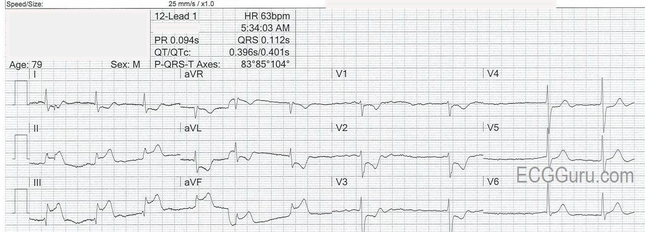
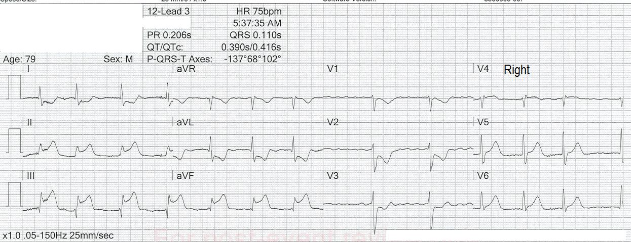
The patient:79-year-old man complaining of severe “burning” chest pain, radiating to his neck. Walking exacerbates his discomfort.He has had nausea and vomiting for 24 hours. Past medical Hx includes high cholesterol and atrial fibrillation. Medications not known.
The ECGs:These ECGs could be called “classic”.There is a 100% occlusion of the right coronary artery (RCA), which was successfully repaired in the cath lab.About 80% of inferior wall M.I.s are due to occlusion of the right coronary artery.Depending on how proximal the occlusion is, we can expect a pattern on the ECG representing injury to all areas supplied by the RCA.This “package deal” can include:
At the ECG Guru website, our main goal is to provide quality teaching materials to those who teach ECG interpretation and other cardiac topics.This ECG offers teaching opportunities for those who teach any level of student.
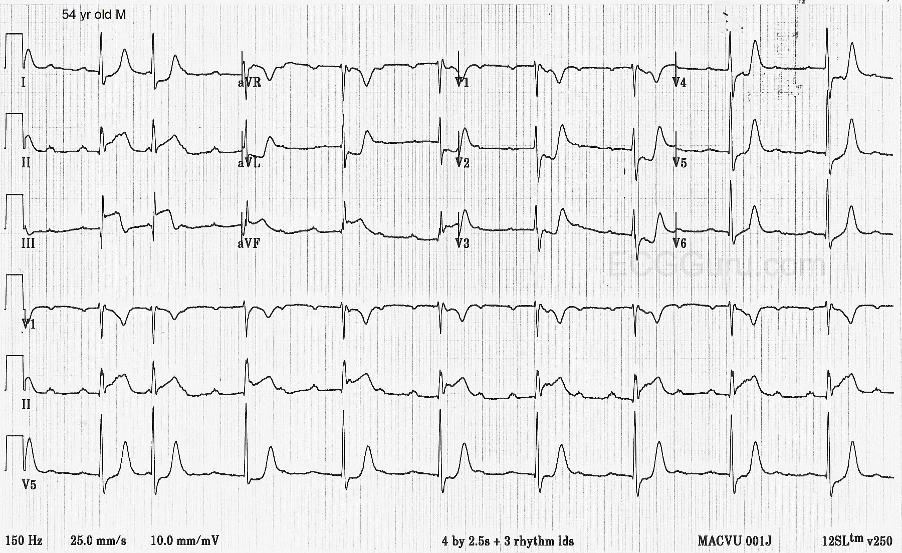
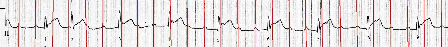
The patient:This ECG was obtained in the Emergency Department from a 54-year-old man who was complaining of severe chest pain and nausea.His BP was 130/68.