Displaying 1 - 4 of 4
Dawn's picture

Normal ECG In A Young Adult

This ECG was obtained from a 24-year-old man who was seen in the Emergency Department for chest pain that was determined to be non-cardiac in origin.  He had a fever and cough, with pain on inspiration. His vital signs were within normal range, and he appeared well-perfused. There was no complaint of dizziness or syncope.

So, what does his ECG show?  The ECG should be interpreted in the context of the age and presentation of the patient.  He is young, and has been healthy all his life.  He is lean and reasonably fit.

The rhythm:  the rate is 81 bpm, and the rhythm is regular.  His P waves are upright in Leads I and II, and they are followed by QRS complexes.  The rhythm is NORMAL SINUS RHYTHM.

Intervals:  The PR interval is 137 ms (.137 seconds), and his QRS duration is 91 ms (0.9 seconds).  His QTc is 404 ms.  All are within normal range.

QRS frontal plane axis:  Normal axis, at around 30 degrees.  Lead II has the tallest QRS of the limb leads, which is an indication of axis in the normal range.  When the electrical axis travels towards Lead II, we can expect Lead aVL to be small, or even biphasic.

Dawn's picture

Ask the Expert

Dr. Jerry W. Jones, MD, FACEP, FAAEM

QUESTION:

Dr. Jones, what advice do you have for teaching ECG beginners? 


Today’s expert is Dr. Jerry W. Jones, MD, FACEP, FAAEM
 

Jerry W. Jones, MD FACEP FAAEM is a diplomate of the American Board of Emergency Medicine who has practiced internal medicine and emergency medicine for 35 years. Dr. Jones has been on the teaching faculties of the University of Oklahoma and The University of Texas Medical Branch in Galveston. He is a published author who has also been featured in the New York Times and the Annals of Emergency Medicine for his work in the developing field of telemedicine. He is also a Fellow of the American College of Emergency Physicians and a Fellow of the American Academy of Emergency Medicine and, in addition, a member of the European Society of Emergency Medicine. 

Dr. Jones is the CEO of Medicus of Houston and the principal instructor for the Advanced ECG Interpretation Boot Camp and the Advanced Dysrhythmia Boot Camp. 

ANSWER: 

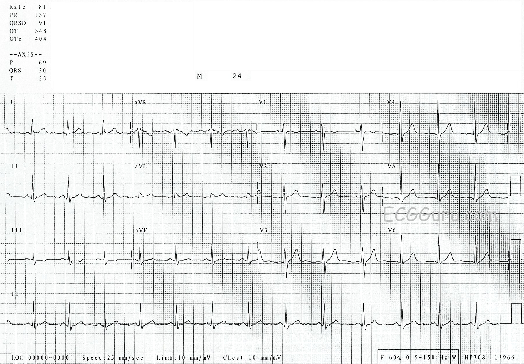
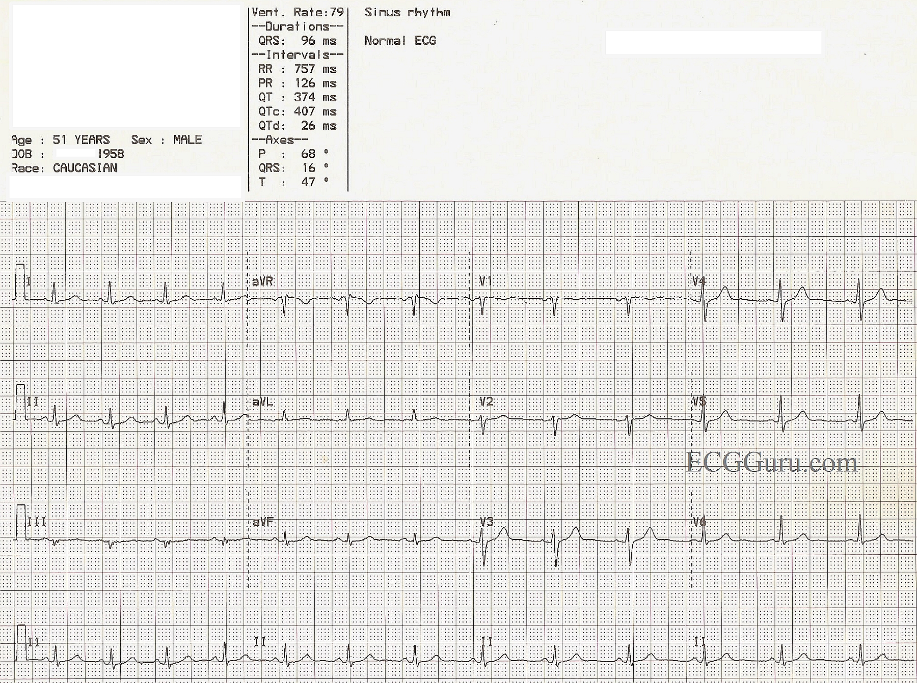
Even in my advanced classes I begin with "normal" ECGs. Throughout my residency in internal medicine, I was never up at 3 am wondering if an ECG was ABNORMAL ... I was always trying to decide if a finding was really NORMAL instead.

Here are a few of my thoughts...

A biphasic P wave in V1 is basically the norm. Even when there is only a monophasic deflection, it's usually because the other half of the biphasic deflection is isoelectric.

In my advanced courses we always begin with a normal tracing and I have all the participants measure the R-R intervals with ECG calipers to demonstrate that there is often considerable variation in the rhythm and that there is very rarely a perfectly regular sinus rhythm (and when there is - it's only for a few moments!). This comes in handy occasionally in deciding whether a tachycardia is sinus or not.

I often find that beginners have the impression that the R waves in the precordial leads increase in size from V1 through V6 - and that should never be the case in a "normal" ECG. Typically the tallest R wave peaks at V4 or V5. Because the V6 electrode is the furthest of all the regular precordial leads from the surface of the heart, it actually diminishes in amplitude. When the R wave in V6 is the tallest across the precordium, it means that the heart has enlarged enough to extend its surface a lot closer to the V6 electrode. That alone is a very good indication of cardiac enlargement.

One other thing I would really emphasize to a newbie is that the ST segment should rise gently into the upslope of the T wave and that there should never be a perceptible angle indicating where the ST ends and the T wave begins - it should be smooth and without a discernible margin. And the T wave should always be asymmetrical - NOT symmetrical. However, when the downslope of the T wave returns to the baseline it CAN create a noticeable angle. 

I hope some of these comments help you teach those who are just beginning to read ECGs.

Dawn's picture

Normal ECG

A nice, normal ECG for your collection.  It always helps to have a normal ECG for your students to compare to the abnormal examples you show them.  It pays to teach the characteristics of "normal" to your students so that they might readily recognize "abnormal".  One could argue that the voltage in this ECG is rather low, but that can be explained by body habitus.  Whether you are teaching basic rhythm interpretation, axis determination, R wave progression, or intervals, this ECG can be of help to you.  Remember, if you are teaching students who have not yet learned 12-lead interpretation, the bottom line of a four-channel ECG is usually a rhythm strip - most often Lead II.  It is easy to download this image, then crop it using any photo management program to include only the rhythm strip.  At the ECG Guru, we use Irfanview for the Instructors' Collection ECGs.

Dawn's picture

Normal 12-Lead ECG

Sometimes, when teaching a class, it can actually be a challenge to find a good example of "normal" for your students.  We all tend to collect the ECGs that are "interesting" or unusual.  It is very important for students to fully recognize "normal" before they can appreciate "abnormal".  Here is a nice example of a normal 12-Lead for you. It was obtained by a portable machine, like those used by EMS or on crash carts, so it does not include a fourth channel rhythm strip.  Challenge your students to find the "normal" characteristics of this ECG: normal sinus rhythm; Lead II has the tallest QRS of the limb leads, indicating a normal axis; Lead V1 is primarily negative and the chest leads progress in an orderly fashion to a positive QRS in V6; no ST segment elevation or depression; no T wave inversions; no pathological Q waves; no abnormally tall or wide QRS complexes; and all intervals are normal.  Challenge your students to pick out the normal features of this ECG.


All our content is FREE & COPYRIGHT FREE for non-commercial use

Please be courteous and leave any watermark or author attribution on content you reproduce.