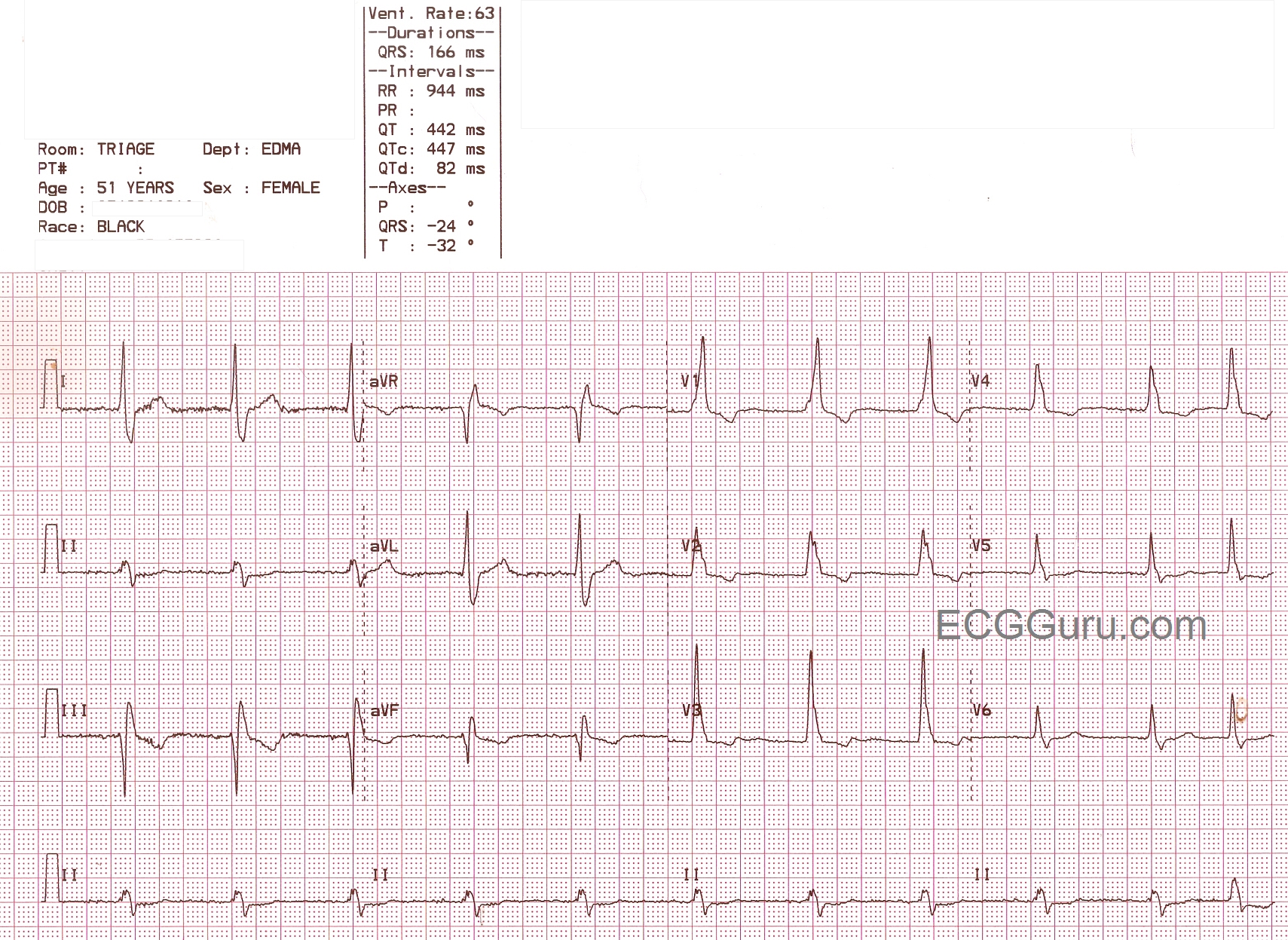
This is quite an interesting ECG, and the ECG Guru would love to hear what you think about it. What we do know is that it is a wide-complex bradycardia in a patient for whom we have no clinical data, except that she is a 51 year old female. The rhythm is probably junctional, as no P waves are seen and the rhythm is regular. The rate of 63 per minute would be consistent with that. Interestingly, no disassociated sinus P waves are seen. All slow wide-complex rhythms should be evaluated for idioventricular origin, or AIVR. The QRS in V1 shows an atypical right bundle branch block pattern. We usually look for rSR', or "bunny ears", but this ECG shows an upright R wave with a smaller, slurred r wave before it. What makes this look like RBBB is the prominent wide little S wave in V6 and in Lead I. We question the R wave progression, too. Do you think it is possible that the electrodes for V2 and V3 are switched? The axis is leftward, causing Lead II to be nearly biphasic - it represents a synthesis of what is seen in Leads I and III. This is enough left axis shift to diagnose a left anterior fascicular block (with RBBB = bifascicular block).
This is a great ECG, and we can't wait to hear from all you ECG Gurus out there. Maybe we will need to adjust our diagnosis after we hear from you.