Displaying 31 - 40 of 42
Dawn's picture

Recent Anterior-Septal Wall M.I. With Right Bundle Branch Block

This is an ECG from a 95 year old man who was recovering from an anterior-septal wall M.I.  Other clinical data for this patient has been lost, except that he suffered a new right bundle branch block during this M.I.  The ECG shows pathological Q waves in V1, V2, and V3, consistent with permanent damage (necrosis) in the anterior septal wall.  The ST segments in those leads are coved upward.  Even though the J points are not elevated, this ST segment shape suggests recent injury.  The classic RBBB pattern is present:  wide QRS, rSR' pattern in V1, and wide little s waves in I and V6.  It is not known why the overall voltage is low in this patient.

Dawn's picture

Normal Sinus Rhythm With Aberrantly-Conducted PACs

This is a normal 12-Lead ECG with two PACs that are aberrantly conducted in a right bundle branch block pattern. (Sixth and ninth beats). In the PACs, the QRS is slightly wider than the normal beats.  The aberrantly-conducted beats have an rsR' pattern in V1, and a wide little S wave in aVL. No PACs are seen in Lead I to demonstrate the wide S wave.  This represents a right bundle branch block pattern, which is a common form of aberrancy, and is rate-related.  That is, the PAC occurs early in the cycle, catching the right bundle branch is a refractory state and unable to depolarize.  Slower beats are easily acommodated by the right bundle branch.

Dawn's picture

Right Bundle Branch Block With Atypical QRS in V1 and LAFB or Ventricular Rhythm???

This is quite an interesting ECG, and the ECG Guru would love to hear what you think about it.  What we do know is that it is a wide-complex bradycardia in a patient for whom we have no clinical data, except that she is a 51 year old female.  The rhythm is probably junctional, as no P waves are seen and the rhythm is regular.  The rate of 63 per minute would be consistent with that. Interestingly, no disassociated sinus P waves are seen.  All slow wide-complex rhythms should be evaluated for idioventricular origin, or AIVR.  The QRS in V1 shows an atypical right bundle branch block pattern.  We usually look for rSR', or "bunny ears",  but  this ECG shows an upright R wave with a smaller, slurred r wave before it.  What makes this look like RBBB is the prominent wide little S wave in V6 and in Lead I.  We question the R wave progression, too.  Do you think it is possible that the electrodes for V2 and V3 are switched?  The axis is leftward, causing Lead II to be nearly biphasic - it represents a synthesis of what is seen in Leads I and III.  This is enough left axis shift to diagnose a left anterior fascicular block (with RBBB = bifascicular block). 

This is a great ECG, and we can't wait to hear from all you ECG Gurus out there. Maybe we will need to adjust our diagnosis after we hear from you.

Dawn's picture

Right Bundle Branch Block With Left Anterior Fascicular Block

This is a nice, clear right bundle branch block pattern: wide QRS, supraventricular rhythm (NSR), and rSR' pattern in V1. Wide little s waves in Leads I and V6 are also diagnostic. The left axis deviation indicates a left anterior fascicular block, since there is no other apparent reason for the left axis deviation, such as pathological Q waves or LVH. Left anterior fascicular block is a diagnosis of exclusion, also considering that RBBB and LAFB are often seen together (bifascicular block), since the two fascicles have the same blood supply.


Dawn's picture

Third-degree AV Block and Junctional Escape Rhythm With Right Bundle Branch Block and Prolonged QTc Interval

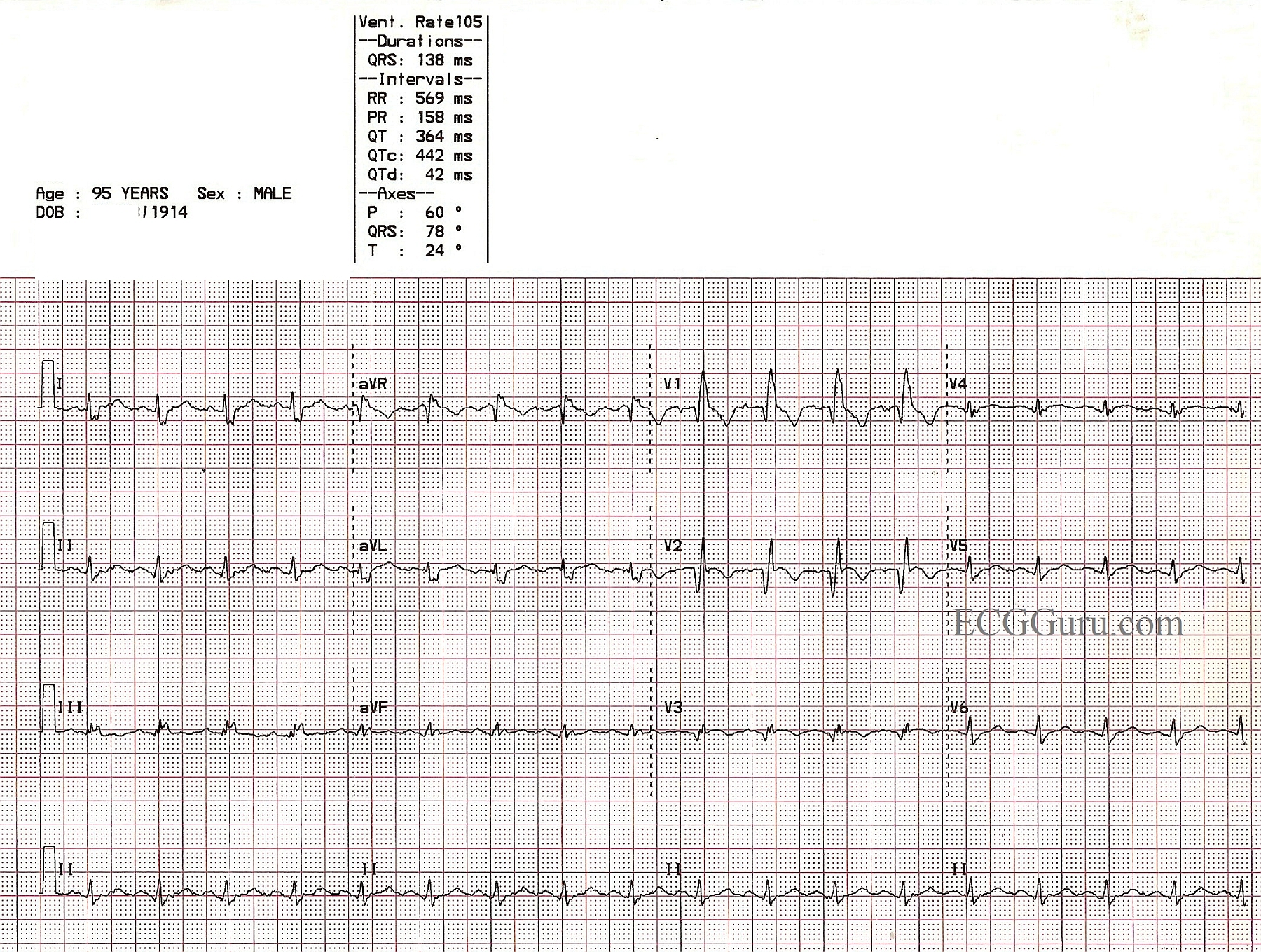
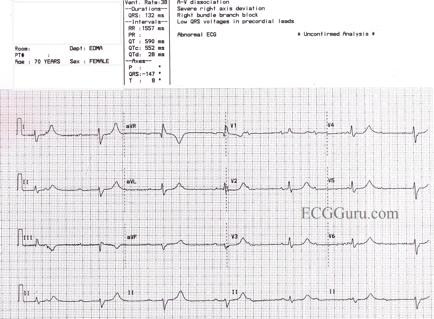
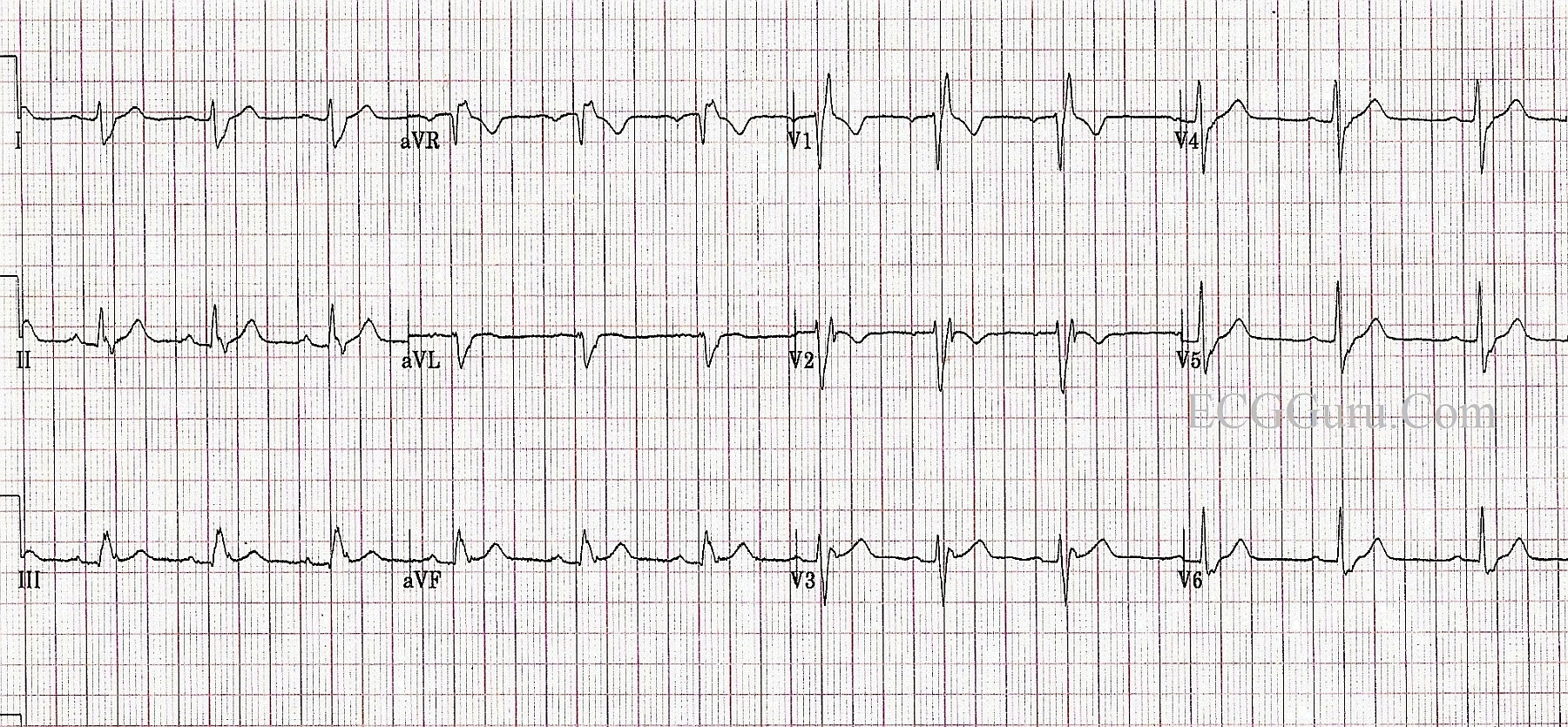
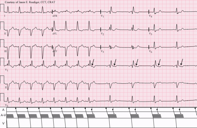
This ECG is from a 70 year old woman for which we have, unfortunately, no clinical information.  It shows a sinus rhythm with a rate of about 72 bpm (NSR) with AV dissociation caused by third-degree heart block.  The escape rhythm is junctional at a rate of 38 bpm.  There appears to be a right bundle branch block, based on the QRS duration of 132 ms, and a wide S wave in Leads I and V6.  The precordial leads do not show the usual RBBB pattern of rSR' in V1 and V2, and the r wave progression is poor (non-existent).  This is felt to be due to poor lead placement (a good teaching point).  Of interest, the ECG machine has reported a "severe right axis deviation" based on the tall upright R wave in aVR and the deep S in avF.  In RBBB, the first part of the QRS represents left ventricular depolarization, and the terminal wave represents the delayed right ventricle.  In effect, the two ventricles have their own electrical axes, which we can see because the ventricles are not depolarizing simultaneously.  The axis of the LV appears to be normal in this tracing.

In addition to the above, this patient has a very prolonged QT interval.  The QT is longer in bradycardic rhythms, but when corrected to a standard of 60 bpm (QTc), this patient's QT interval is still prolonged at QTc: 552 ms.  Without clinical data, we cannot speculate  as to why this patient's QTc is prolonged, but it can be a very dangerous situation.  Follow the links for more information on QT prolongation and Torsades de Pointes and Long QT Syndrome.

As always, we welcome comments from our members adding insight to this interesting ECG, and also questions you would like to ask our Guru members.

Dawn's picture

Right Bundle Branch Block and Previous Anterior Wall M.I.

This example of previous anterior wall M.I. with right bundle branch block is from a 95-year-old man.  He survived an anterior-septal M.I. caused by an occlusion of the left anterior descending branch of the LCA. The septum was involved in the damaged area, and the patient was left with a right bundle branch block.  The pathological Q waves in Leads V1 through V3 show the location of permanent damage, or necrosis.

In this case, the usual rSR' pattern of right bundle branch block is replaced by qR, indicating loss of the initial r wave due to the M.I.

Review of RBBB criteria:  wide QRS, rSR' pattern in V1, and Rs with a slurred s wave in I and V6.  These criteria must be present in a SUPRAVENTRICULAR rhythm to diagnose RBBB.

Dawn's picture

Right Bundle Branch Block

This ECG is from a healthy young man in his 20's.  He was born with a ventricular septal defect (VSD) that was surgically repaired when he was a toddler.  He now has a right bundle branch block, which could be a result of the defect, or the surgery.  This is a good ECG for the Instructors' Collection because it clearly shows all the ECG characteristics of right bundle branch block:  wide QRS in a supraventricular rhythm (in this case, NSR), rSR' pattern in V1, wide or "slurred" S waves in Leads I and V6.  There is no rhythm strip below the 12 leads in this ECG, but there is no rhythm disturbance.

jer5150's picture

Jason's Blog: ECG Challenge of the Week for Oct. 21-28, 2012

No clinical patient data available for this 12-lead ECG.

What does this tracing show?  Choose the correct answer from the list below.

(1.)  Sinus bradycardia with atrial bigeminy; conducted APBs; prominent U-waves; RBBB
(2.)  Sinus rhythm with 3:2 and 2:1 Type II AV block; RBBB
(3.)  Sinus rhythm with atrial bigeminy; both conducted and nonconducted APBs; RBBB

Acronyms:
APBs = atrial premature beats
RBBB = right bundle-branch block

Dawn's picture

Anterior Wall M.I. With Bifascicular Block

This is a good example of acute anterior wall M.I., with ST elevation in V1 through V6, as well as in Leads I and aVL.  The extensive distribution of ST segment elevations across the anterior and high lateral walls indicates a proximal LAD artery occlusion.  In addition, this ECG shows right bundle branch block, with a QRS width of 144 ms (.14 sec.) and an rsR' pattern in V1. There is also a wide s wave in Lead I which is partly obscurred in V6 by the ST elevation.  The right axis deviation (98 degrees) suggests a left posterior fascicular block which, when coupled with the RBBB, is a bi-fascicular block.  P waves are difficult to see.  Do you think they are found at the end of the QRS complexes, representing a long first-degree AVB?  Look at leads V3 through V6 for clues.

Please feel free to add your comments below.  The more "gurus" the better.

A good ECG to teach your students that a patient facing a life-threatening emergency may have a "normal" rate and regular rhythm.  There is something in this ECG for beginners through advanced students.

Dawn's picture

Sinus Rhythm With Non-Conducted Atrial Bigeminy

Jason's Blog: ECG Challenge of the Week, 6-8-12     INTERPRETATION:


1)  In first half of strip: Normal sinus rhythm (rate = 100/min) with . . .

2)  . . . bifascicular block—right bundle-branch block plus left anterior hemiblock 

      (RBBB + LAHB), left axis deviation (LAD) at -57 degrees.

3)   In second half of strip: Sinus rhythm interrupted by a run of nonconducted atrial bigeminy  (arrows (↓); see laddergram).


Jason E. Roediger, CCT, CRAT

Pages

All our content is FREE & COPYRIGHT FREE for non-commercial use

Please be courteous and leave any watermark or author attribution on content you reproduce.