Displaying 1 - 3 of 3
Dr A Röschl's picture

Long QT Syndrome,Look Closely and Do Not Trust the ECG-Computer

ST-elevation V2/V3, why? The patient never had chest pain, echocardiography inconspicious. History of several syncopal episodes. But, there is something wrong with the ECG. We observe a sinus rhythm ECG, at first glance, there are ST-elevations in leads V2-V4. No ST-depressions are visible. Upon closer examination, a prominent T-wave is noted, starting immediately after the QRS complex. The ECG computer erroneously indicates a significantly shortened QT and QTc interval.

Dawn's picture

Incorrect Machine Interpretation

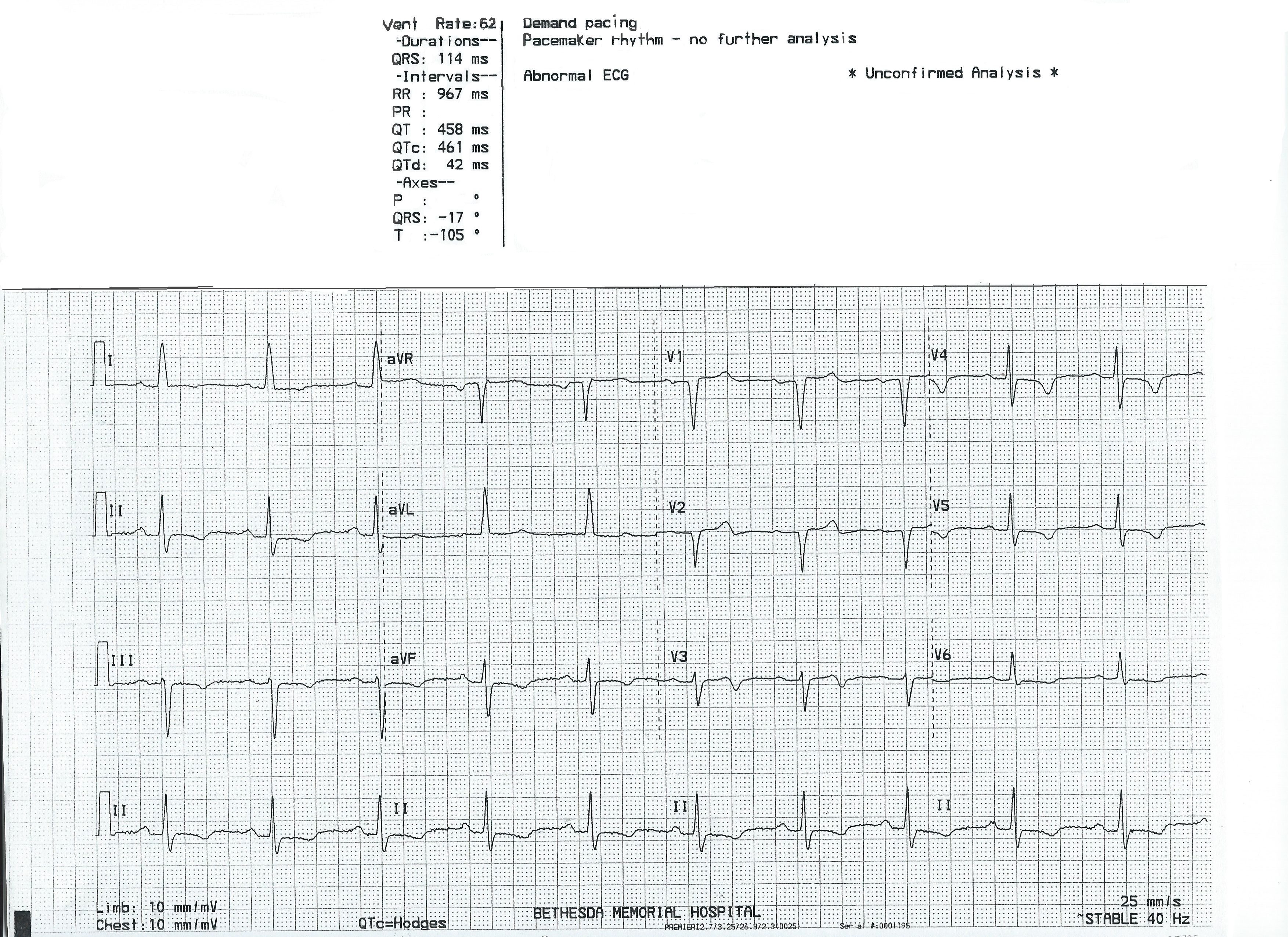
This ECG is presented as an example of INCORRECT MACHINE INTERPRETATION.  While there are many abnormalities in this ECG, it does not represent a paced rhythm. While there are exceptions, most paced rhythms represent either AV sequential pacing, right ventricular pacing, or bi-ventricular pacing.

RECOGNITION OF A PACED RHYTHM

Recognizing a paced rhythm can be difficult in some cases. Because pacemakers now have so many programmable features, there is a wide variety of ECG changes associated with them.  Pacer “spikes” can be difficult to see in all leads.  Finding evidence of the device on the patient’s chest or via patient history is a big help in reminding us to scrutinize the ECG for paced rhythm.

An AV sequential pacemaker or a right ventricular pacemaker will pace the ventricles via the right ventricle.  This produces a WIDE QRS and a leftward axis, often causing Leads II, III, and aVF to be negative and aVL and aVR to be positive.  Along with the wide QRS, we will see DISCORDANT ST CHANGES.  That is, there will be ST depression and T wave inversion in leads with positive QRS complexes and ST elevation and upright T waves in leads with negative QRS complexes.

Bi-ventricular pacing can be a little more complicated to recognize, as the QRS can be narrow, with signs of fusion between the wave produced by the LV electrode and the RV electrode.

The frontal plane axis is usually far right – aVR will be positive.  Lead I will be negative.

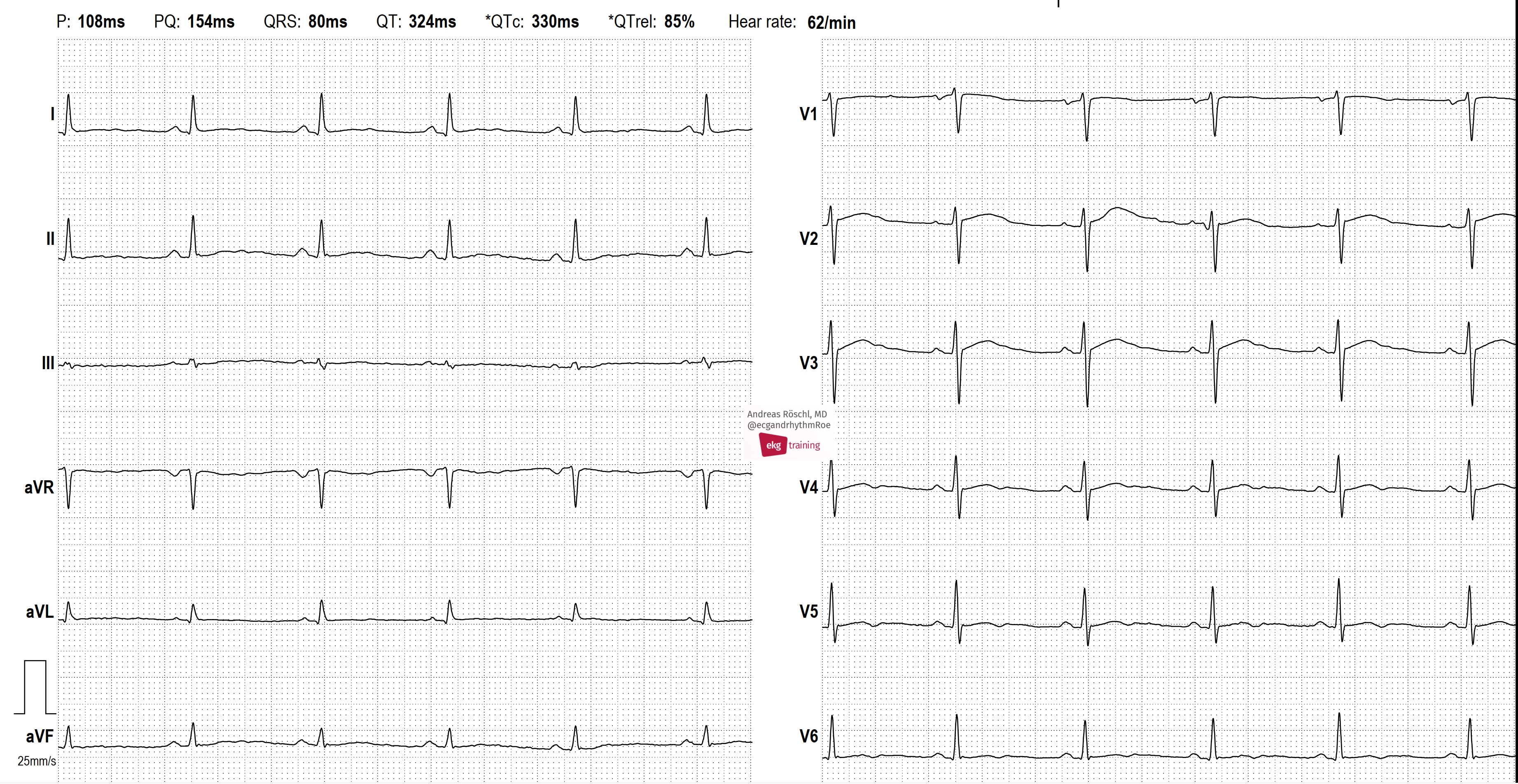
The machine is wrong:  there is no indication of a pacemaker, and P waves are present, even though they are not noted in the "PR Interval" or "P Axis".

SO, THIS IS NOT A PACED RHYTHM – WHAT IS IT?

Dawn's picture

Complete AV Block With Junctional Escape Rhythm

This ECG is from a 78-year-old woman.  We do not know any clinical details.

 We break from our usual habit of removing the ECG machine’s interpretation of the ECG to serve as a reminder that the computer interpretation can be wrong.  ECGs should ALWAYS be interpreted by a knowledgeable person.  The machine interpretation can serve as a reminder, but should not take the place of human interpretation. 

Here is what we DO see:  There is a normal sinus rhythm present, as evidenced by the regular P waves that do not change their morphology.  Some of the P waves are “buried” behind QRS or T waves.  The atrial rate is 95 bpm. 

The ventricular rhythm, at 40 bpm, is also regular, but is separate from the atrial rhythm.  Even though some of the P waves LOOK like they have conducted to produce QRS complexes, they have not.  The PRIs are not all the same.  Neither do they “progressively prolong”.  There is no irregularity of the QRS rhythm or variation in QRS morphology.  We see the classic “AV DISSOCIATION” of complete heart block. 

When there is a third-degree AV block with a narrow-QRS escape rhythm, we can assume the block is in the AV node.  The junction is the escape focus, producing a narrow-complex rhythm between approximately 40-60 bpm.  In this case, the QRS is slightly wide at 112 ms (.11 sec), and the QRS complexes in several leads are fragmented.  Some might argue that there is an idioventricular escape mechanism.  But, with a normal frontal plane axis, borderline width,  and no T wave inversions, the rhythm looks more supraventricular.  The R wave progresson on the precordial leads shows a persistently negative QRS with late transition in V5.  The QRS complexes in V1 and V2 appear to have pathological Q waves.  When R wave progression is not normal, we should also consider electrode misplacement. 

All our content is FREE & COPYRIGHT FREE for non-commercial use

Please be courteous and leave any watermark or author attribution on content you reproduce.