Displaying 11 - 20 of 42
Dawn's picture

Bifascicular Block With First-degree AVB

The Patient  This ECG was obtained from an 80-year-old man with a past medical history of hypertension, diabetes, chronic obstructive pulmonary disease, hyperlipidemia, chronic kidney disease, and heart failure with preserved ejection fraction (HFpEF).


He presented to the hospital with a complaint of shortness of breath, and was determined to have an exacerbation of his COPD in the setting of a viral infection.  He was treated with breathing treatments and steroids, and was discharged home in improved condition.

The ECG   There is a sinus rhythm at 80 bpm.  The QRS complex is wide at .18 seconds (180 ms). The PR interval is slightly prolonged at .24 seconds (240 ms), which is first-degree AV block. There is right bundle branch block (QR in V1 and rS in Leads I and V6 with a wide QRS).  There is also a left posterior fascicular block (also called hemiblock), recognized by the right axis deviation (III is taller than aVF ).  This is bi-fascicular block, which can be chronic or acute.  There is very slight ST elevation with flattening in V1 and possibly V2.  In RBBB, “normal” T waves are directed opposite the terminal wave of the QRS.  V3 should, then, have inverted T waves. So, the upright T wave in this patient could be considered to be equivalent to an inversion in a normal ECG.  V4 through V6 have biphasic T waves, which is not normal for RBBB.  Because we do not have an old ECG, we don’t know which, if any, of these changes are chronic.  With this patient’s past medical history, it is possible for any of them to be pre-existing.

I will admit that, when I first saw this ECG, the V1 rhythm strip at the bottom looked as though there were regular P waves at a rate of about 220-240, with one buried in each QRS and one in each T wave. In scrutinizing all the other leads, I cannot find evidence for an underlying atrial tachycardia or atrial flutter, so it is probably a coincidence that the P and T “march out” with the assumption of a hidden P wave in the QRS.

Dawn's picture

Bifascicular Block

This ECG is from a 77 year old woman who was brought to the Emergency Department by EMS. She was found to be suffering from sepsis.

ECG Interpretation      The ECG shows the expected sinus tachycardia at 123 beats per minute.  There is significant baseline artifact, of the type usually seen with muscle tension.  The artifact makes it difficult to assess P waves and PR intervals.


What we do see is RIGHT BUNDLE BRANCH BLOCK and LEFT ANTERIOR HEMIBLOCK, also called LEFT ANTERIOR FASCICULAR BLOCK.  Together, these are called BIFASCICULAR BLOCK.  Most people have three main fascicles in the interventricular conduction system:  the right bundle branch and the two branches of the left bundle branch, the anterior-superior fascicle and the posterior-inferior fascicle.  In bifascicular block, two of the three are blocked.

The ECG criteria for right bundle branch block are:

     *     wide QRS (> .12 seconds)


     *     rSR’ pattern in V1 .  (the initial R wave may be hard to see, but the QRS will be predominantly upright.

Dawn's picture

Right Bundle Branch Block With Probable Previous M.I.

This ECG was obtained from an 87-year-old man with chest discomfort.  We have no other clinical information.

ECG Interpretation   The rhythm is regular and fast, with P waves, at 95 beats per minute. So, it is normal sinus rhythm, but the rate is probably not “normal” for this patient.  The P waves are small, and difficult to see.  We suggest Lead I to best view the P waves in this example. This is a good opportunity to teach the value of evaluating rhythm strips in more than one simultaneous lead, as subtle features may not show up well in all leads.  There is a first-degree AV block, with a PR interval of 232 ms.

We see the right bundle branch block (RBBB) pattern: rSR’ in the right precordial leads (with a tiny q wave in V1, which is not typical of  RBBB).  The QRS is wide at 148 ms (.148 seconds).  The R prime (R’) represents the right ventricle depolarizing slightly after the left ventricle.  This terminal delay widens the QRS without affecting the depolarization or contraction of the left ventricle.  This delay can be seen in every lead, but is especially easy to see in Leads I and V6, where there is a wide little s wave.  It is normal for the T waves to be in a direction opposite that of the terminal wave (inverted in Leads V1 and III, for example.)

There is left axis deviation.  The causes of LAD are many.  It is not unusual for people with RBBB to also have a left anterior hemiblock (LAH), also called left anterior fascicular block.  The left anterior fascicle has the same blood supply as the right bundle branch.   LAH causes a frontal plane axis shift – instead of Lead II having the tallest QRS of the limb leads, Leads I and aVL will be the tallest upright QRS complexes of the six limb leads.  Lead II will be very small, or flat, or negative. However, the probability of pathological Q waves in the inferior leads offers a more likely explanation for the leftward axis shift.  The M.I. that would have caused these Q waves is old, as there are no acute ST changes.  It would, of course, help to know this patient’s history.

Right bundle branch block can make evaluating for ST segment elevation a bit tricky.  Occasionally, the terminal delay – especially in Leads III and aVF – can be mistaken for ST elevation.  The J points in this ECG all appear to be at the baseline, with no overt STEMI.


Dawn's picture

Acute M.I. With Right Bundle Branch Block and Atrial Pacing

This ECG was taken from a 78-year-old man who was experiencing chest pressure in the morning, after having left shoulder pain since the night before. He has a history of hypertension and hypercholesterolemia, and has an implanted pacemaker.

What does the ECG show?  The ECG shows an atrial paced rhythm, with two premature beats, beats number 5 and 12.  These are probably PVCs.  The patient has a functioning AV conduction system, so the paced atrial beats are conducting through the AV node and producing QRS complexes.  In the interventricular conduction system, the impulse encounters right bundle branch block. This causes each QRS to have an “extra” wave attached at the end, representing slightly delayed depolarization of the right ventricle.  Instead of an “rS” pattern in V1, for example, we see “rSR’ “.  The slight delay causes the QRS to be widened, as we are measuring the two ventricles separately, rather than synchronously.

There is definite ST segment elevation in V2 and V3, and the shape of the ST segment is straight, having lost it’s normal “concave upward” appearance.  In an ECG taken three minutes later, the STE extends to V4.

Do the pacemaker or the right bundle branch block prevent us from diagnosing an ST-elevation M.I.?  The answer to that is a resounding “NO!” Pacemakers can sometimes make it difficult to assess ST elevation because ventricular pacing causes ST segment changes.  Pacing the right ventricle causes a depolarization delay in the left ventricle as the impulse travels “cell to cell” across the LV.  This means an RV-paced beat will resemble a PVC from the RV.  When LV depolarization is altered, repolarization will also be altered, causing ST elevation in leads with negative QRS complexes, and ST depression is leads with upright QRSs. These are called discordant ST changes. These changes are proportionate to the height or depth of the QRS, with very minimal or no ST changes in leads with short or biphasic QRS complexes.  We don’t have to worry about that in this situation – the pacemaker is not pacing the ventricles.

Dawn's picture

Right Bundle Branch Block With Machine Interpretation Error

Today’s ECG is from a 74-year-old man for whom we have no clinical information.  It shows a “classic” right bundle branch block.  It also shows an example of the ECG machine getting some of the interpretation wrong.  An early mistake in the interpretative algorithm caused a cascade of inaccuracies.

REVIEW of RIGHT BUNDLE BRANCH BLOCK ECG CRITERIA

     *   Supraventricular rhythm

     *   QRS .12 seconds (120 ms) in width

     *   rSR’ pattern in V1

     *   Small, wide S wave in Leads I and V6

In right bundle branch block, the initial part of each QRS complex represents the depolarization of the septum and left ventricle.  The right ventricle depolarizes late, and is represented by a terminal wave at the end of each QRS.  In V1, that terminal wave is the R’ and in I and V6 it is the small S wave. 

MACHINE MISTAKES  The first mistake the machine made was in measuring the QRS width. The machine says the QRS is .096 seconds (96 ms).  It is actually about .16 - .18 seconds.  Look at the second QRS in V1, and you will see that it extends almost the full width of a wide block (.20 sec).  It is apparent that the machine measured only the left ventricular portion of the QRS complex. Because of this error, the right bundle branch block was not noted. 

The mistake in measuring the QRS complex resulted in the machine misinterpreting the terminal wave as the ST segment.  This resulted in notations in capital letters warning of ST elevation and presence of myocardial ischemia.  The j points are actually at the baseline in all leads, indicating NO ST elevation. 

ST and T WAVE CHARACTERISTICS OF RBBB  Typically, in RBBB, the T wave will be opposite in direction from the terminal (RV) deflection.  So, when there is an R’, there will be T wave inversion.  The j point of the ST segment will not be altered, as the ST segment reflects what is happening in the LEFT VENTRICLE, which is depolarizing normally.  That means that an acute ST elevation M.I. will look the same in RBBB as it does without BBB. 

Dawn's picture

Ask The Expert

Today’s expert is Dr. Jerry W. Jones, MD, FACEP, FAAEM

Jerry W. Jones, MD FACEP FAAEM is a diplomate of the American Board of Emergency Medicine who has practiced internal medicine and emergency medicine for 35 years. Dr. Jerry JonesDr. Jones has been on the teaching faculties of the University of Oklahoma and The University of Texas Medical Branch in Galveston. He is a published author who has also been featured in the New York Times and the Annals of Emergency Medicine for his work in the developing field of telemedicine. He is also a Fellow of the American College of Emergency Physicians and a Fellow of the American Academy of Emergency Medicine and, in addition, a member of the European Society of Emergency Medicine. 

 Dr. Jones is the CEO of Medicus of Houston and the principal instructor for the Advanced ECG Interpretation Boot Camp and the Advanced Dysrhythmia Boot Camp. 


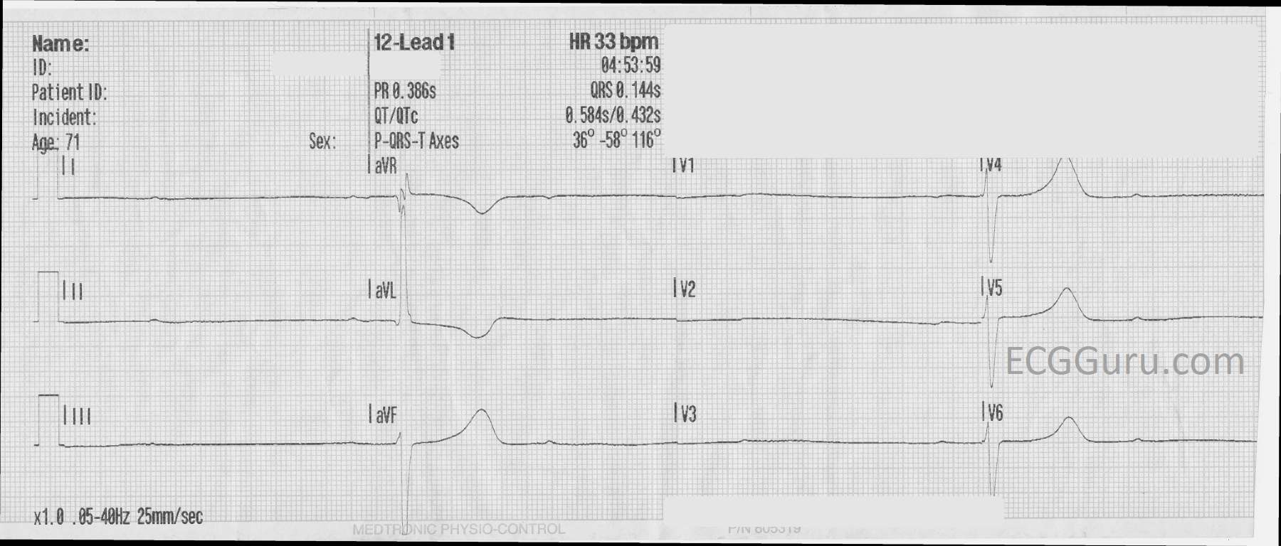
Question:   What is the cause of an apparent right bundle branch block pattern in a paced rhythm?

Answer:  Is There a Pacemaker Wire Problem… or Not?

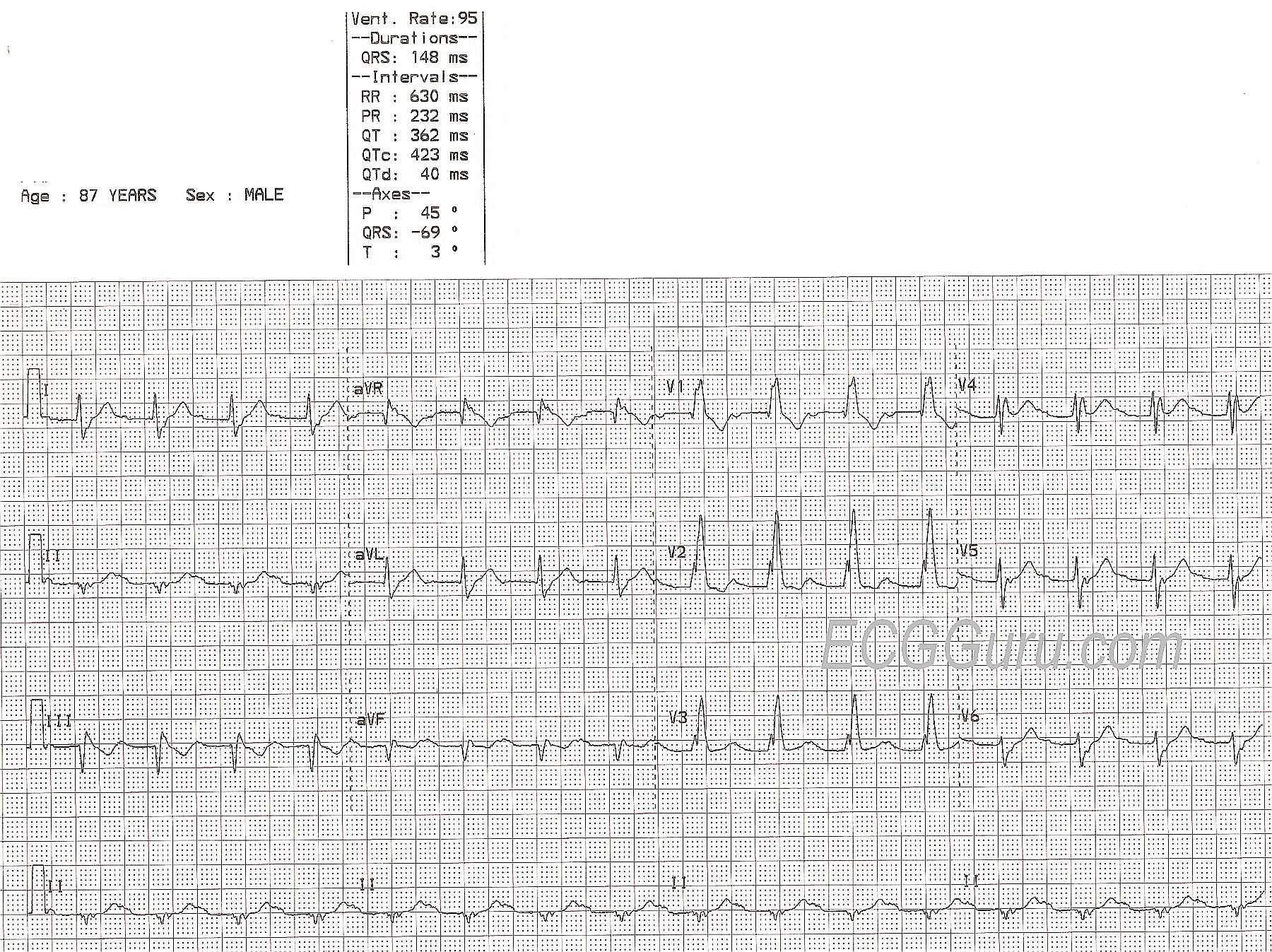
 During one of my orientations as a young internal medicine house officer, the cardiologist lectured to us on the essentials of how to check pacemakers. Since none of us had any ECG interpretation background our comprehension was less than sterling. But I remember him stressing the point that a properly paced pacemaker lead would result in a left bundle branch block pattern on the ECG. A right bundle branch block pattern in V1, on the other hand, meant that the pacemaker wire had inadvertently wandered into the left ventricle – a highly undesirable situation. 

“Not to worry,” he said. “Such things rarely happen and you will probably retire before seeing such a thing!” That evening I saw my first pacemaker 12-lead ECG with a right bundle branch block pattern in V1. Fate wasted no time with me.

I ordered a 3-view chest x-ray and as far as I could see, the wire looked like it was in the right ventricle where it was supposed to be. I called the cardiologist on-call who happened to be in the hospital at the time and he dropped by the ward. Back then, we didn’t have ultrasound or echo available. But he, too, was convinced the pacemaker wire was in the right ventricle. It really was and so I still hadn’t seen a RBBB pattern due to a pacer wire in the left ventricle. I still haven’t, but I have seen a number of pacemaker ECGs with a RBBB pattern in V1.

How do we know if such a finding represents a real left ventricular pacer wire or a pseudo-malplacement?

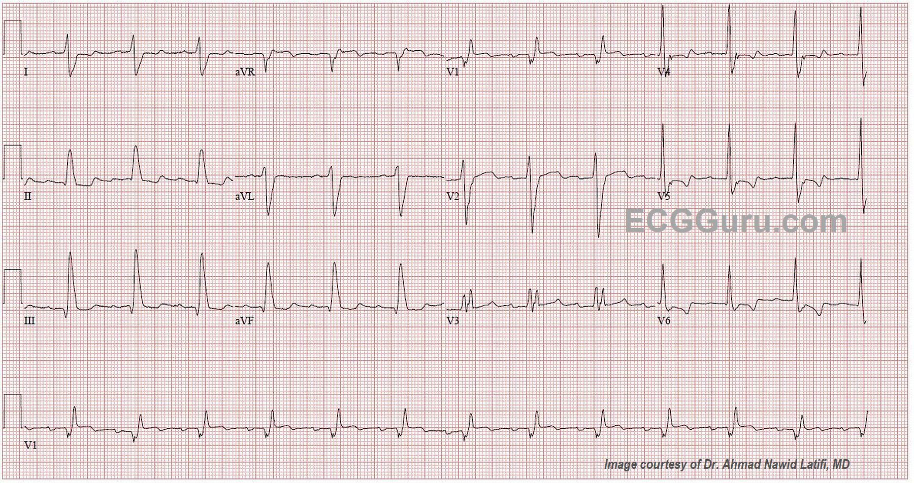
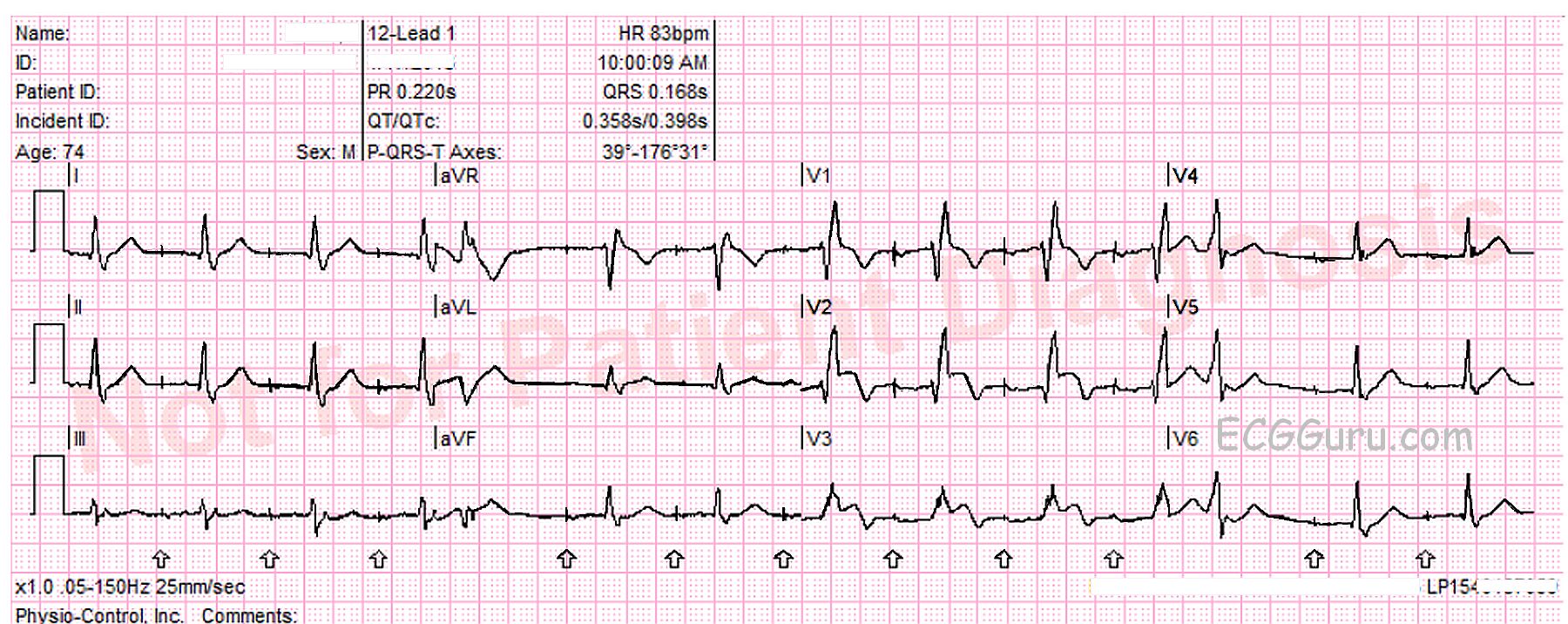
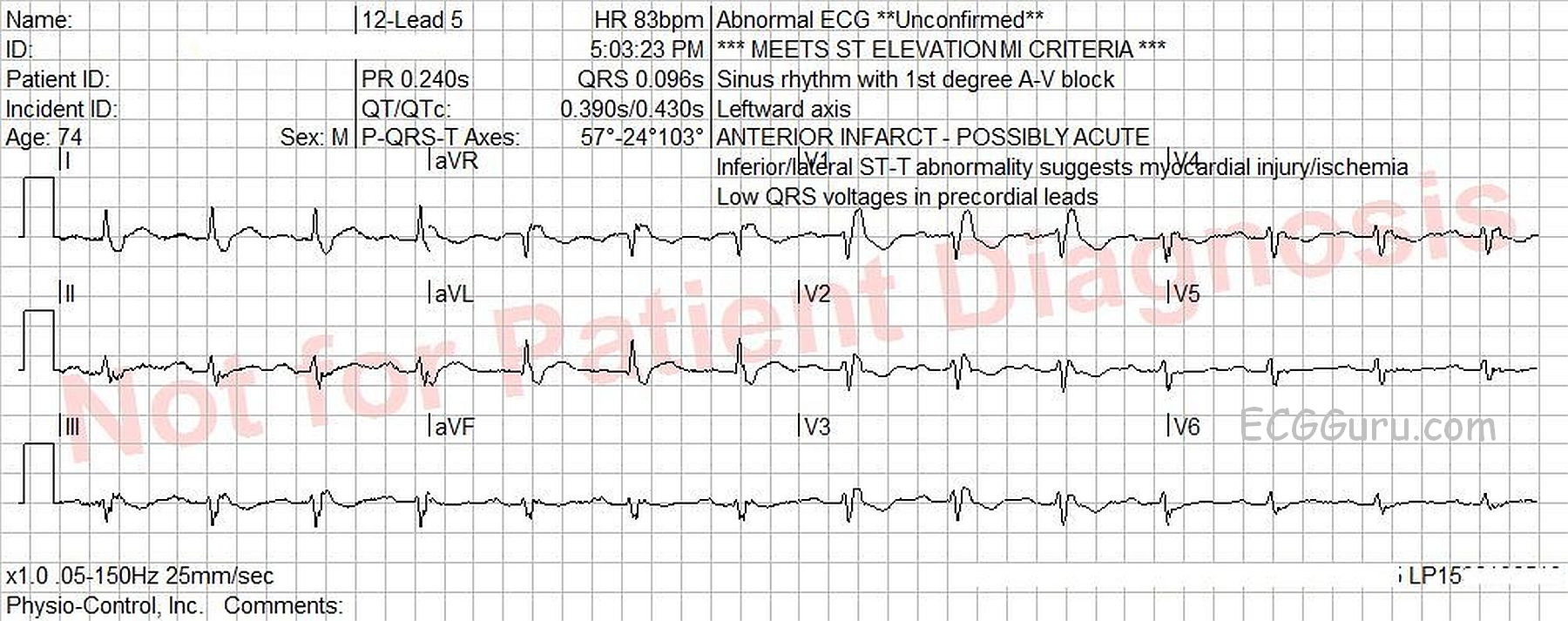
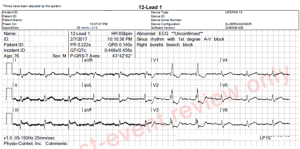
First, just be aware that a wire that really IS in the left ventricle is going to present with a RBBB pattern in V1. It will NOT ever present with a LBBB pattern. However, a wire that has been correctly placed in the RIGHT ventricle can – from time to time – present with a RBBB pattern in V1. In my years as an attending in the emergency department, I saw this seven or eight times.

Second, the axis of the pseudo-malplacement tends to demonstrate a significant left axis deviation, between -30 ° and    -90 °. Since the right ventricle is activated first, the vector finishes by pointing up and to the left. If the wire were actually located in the left ventricle, the mean frontal axis would be to the right of +90 °

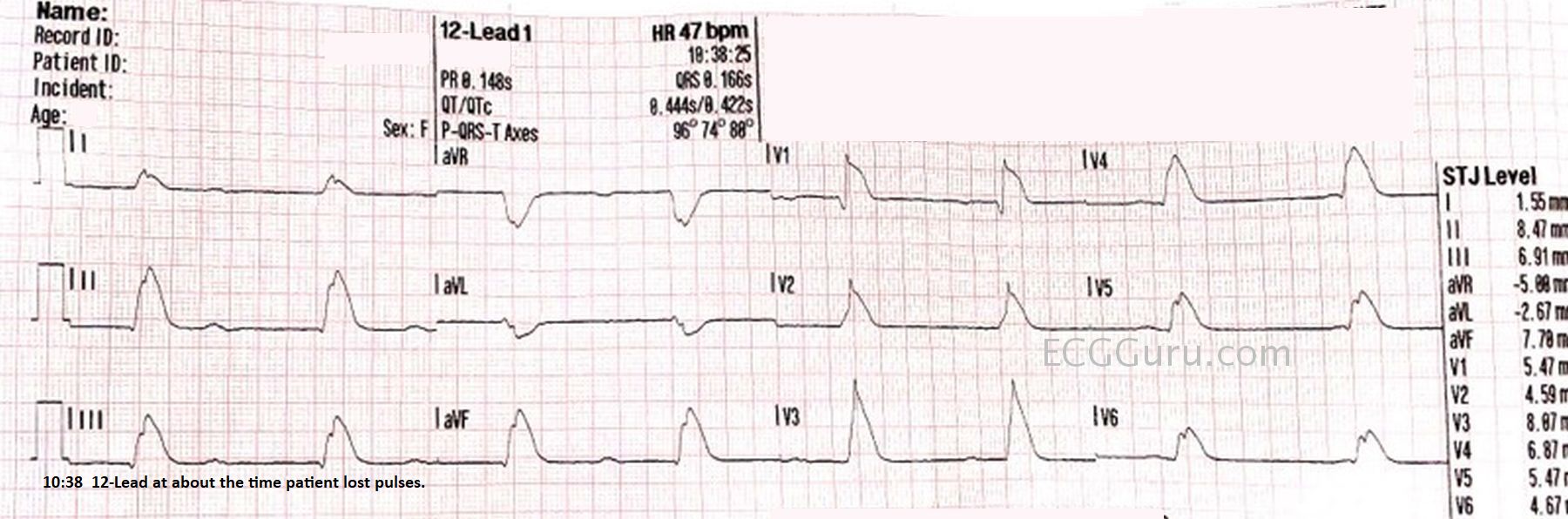
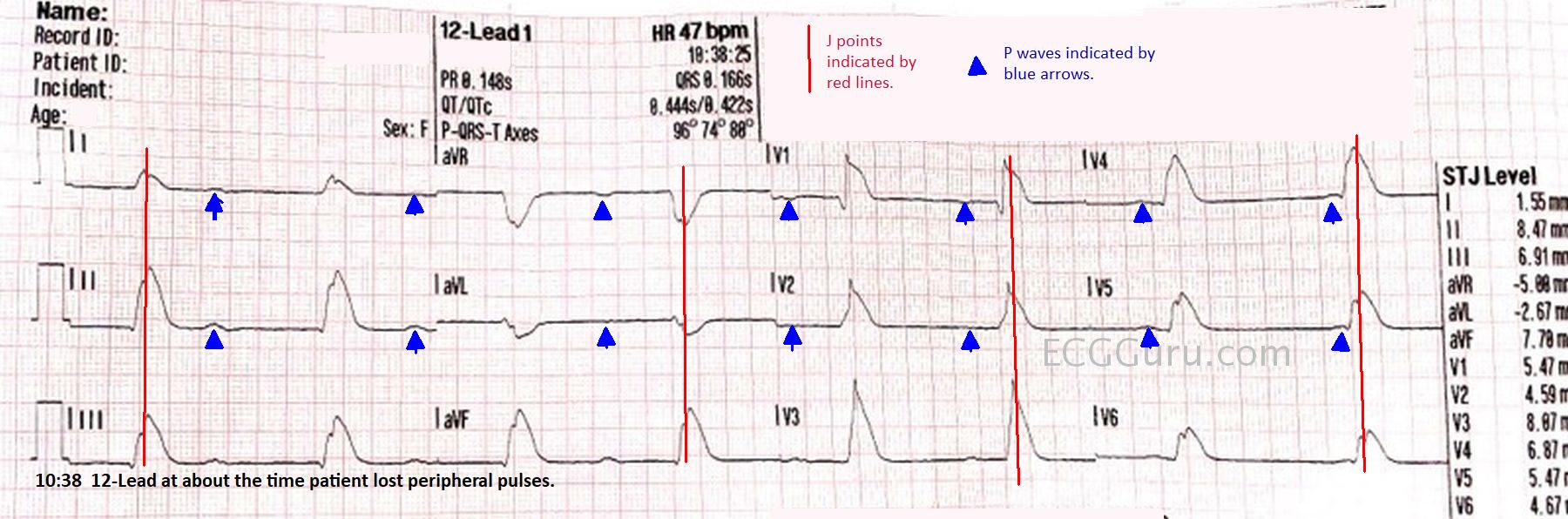
Third, when we look in the precordial leads, we know that Leads V1 and V2 overlie the right ventricle and leads V5 and V6 overlie the left ventricle. Leads V3 and V4 are in between. If the pacemaker wire is in the right ventricle, whatever is causing it to have an RBBB pattern in V1 will disappear before V3. A pacemaker wire in the right ventricle will show a LBBB pattern (QS) by Lead V3. If the wire is truly in the left ventricle, the RBBB pattern will extend to V3 and usually beyond. So a quick check is this: if you see a RBBB pattern in V1 in a pacemaker patient, look at V3. If the RBBB pattern is in V3 also, the wire is truly in the left ventricle. If V3 has a predominately negative QRS (QS), the wire is safely in the right ventricle where it is supposed to be. 

A fourth check is to look for an S wave in Lead I. Remember: one of the most characteristic features of RBBB is that slurred S wave in Lead I (as well as the other left-sided leads). If the ECG shows an RBBB pattern in V1 and an S wave is present in Lead I, then that is most likely a real RBBB pattern and the wire has somehow made its way into the left ventricle.

Pseudo Malplacement of Pacemaker Wire


Dawn's picture

Inferior Wall M.I. and Right Bundle Branch Block

These ECGs were taken from a 76 year-old-man who was complaining of chest pressure for 20 minutes.  He had a remote history of coronary artery bypass graft surgery.

This case has several good teaching points, including:

Significant artifact.  The limb leads show artifact which is severe enough to hamper our assessment of the j point location. Every effort should be made to eliminate artifact.  Some measures that might help are:

        *  clean and slightly "rough up" the skin where the electrode will be placed.  A rough wash cloth or gauze pad will work.

        *  shave hair if necessary.

        *  avoid areas of movement if possible.  Precordial electrodes must be placed in specific spots, but limb leads may be placed anywhere on the limb or on the trunk if it is impossible to avoid movement on the limbs.

       *   use fresh electrodes that have been protected from drying out.

Subtle STEMI changes.   This patient has an inferior wall M.I., which was confirmed as a complete occlusion of the right coronary artery in the cath lab.  The ST elevation in Leads II, III, and aVF is subtle, and more difficult to measure because of the artifact.  However, the SHAPE of the ST segments is a giveaway - they are very straight.  A convex-upward shape is normal (see Lead I).  Also, Lead aVL shows typical ST DEPRESSION, as a reciprocal view of the STE in Lead III.  More ST depressions can be seen in Leads V1 through V3, and they end abruptly there.  These localized ST depressions represent a reciprocal view of the posterior (also called lateral) wall, and represent an "extension" of the inferior wall M.I. up the back of the heart.  A V4 Right lead was obtained and shows no measurable ST elevation, but the shape is straight to slightly "frowning", indicating that the right ventricle may soon have STE.  Repeat ECGs should be obtained to watch for more definite ST elevations.

Dawn's picture

Anterior Wall M.I. With Bifascicular Block

This ECG is taken from an 82-year-old man who called 911 because of chest pain.  He has an unspecified “cardiac” history, but we do not know the specifics. 

WHAT IS THE RHYTHM?  The heart rate is 69 bpm, and there are P waves before every QRS complex. The underlying rhythm is regular, with one premature beat that is wide without a P wave.  The PR interval is slightly prolonged at .25 seconds.  The rhythm is normal sinus rhythm with first-degree AV block and one PVC. 

WHY THE WIDE QRS?   The QRS complex is wide at .14 seconds. The QRS in V 1 has a wide R wave after a small Q wave.  This in consistent with right bundle branch block pattern, with loss of the normal initial small r wave (pathological Q waves).  The diagnosis of RBBB is further corroborated by the wide little S waves in Leads I and V6.  The QRS frontal plane axis is -66 degrees per the machine, and clearly “abnormal left” because the QRS in Lead II is negative, while the QRS in Leads I and aVL are positive.  This is left anterior fascicular block, also called left anterior hemiblock.  The combination of RBBB and LAFB is a common one, as the two branches have the same blood supply.  It is also called bi-fascicular block. 

WHAT ABOUT THE ST SEGMENTS?  The ST segments in leads V2 through V6 are elevated, and their shape is very straight, as opposed to the normal shape of coved upward (smile). Even though the amount of ST elevation at the J points appears subtle, the shape of the segments, the fact that they appear in related leads, and the fact that the patient is an elderly male with chest pain all point to the diagnosis of ANTERIOR WALL ST elevation M.I. (STEMI).  Additional ST changes include a straight shape in Leads I and aVL and ST depression in V1 and aVR.  

PATIENT OUTCOME  The patient was transported to a cardiac center, where he received angioplasty in the cath lab.  The left coronary artery was found to be occluded, and was repaired and stented.  He recovered without complications and was sent home in a few days.

Dawn's picture

Second-degree AV Block, Type II?

This ECG is taken from an elderly woman who complains of feeling weak and tired. We have no other clinical information, unfortunately.

There is an obvious bradycardia, with more P waves than QRS complexes.  Here is what we see:

*  Atrial rate is around 115/min. and P waves are regular and all alike.

*  Ventricular rate is around 35/min. and QRS complexes are regular and all alike.

*  PR intervals, when they occur, are all the same at 162 ms.

*  QRS duration is wide at 122 ms.

*  QTc interval is prolonged at 549 ms.

What does this mean?  There is sinus tachycardia with second-degree AV block because the atrial rate is over 100/min, but not all P waves are conducted.  The AV block looks like a Type II (Mobitz II) block because the PR intervals are all the same.  This is a reliable indicator of conduction. (Not third-degree AVB).  The wide QRS complexes are due to right bundle branch block.  The ECG signs of RBBB are: 1) wide QRS; 2) supraventricular rhythm; and 3) rSR’ pattern in V1 and Rs, with a wide little s wave, in Leads I and V6.

Dawn's picture

Bifascicular Block and Sinus Bradycardia

Today’s ECG is from a 75 year old man who has been experiencing syncope. 

Examination of the ECG shows a sinus bradycardia at just under 40 bpm.  There is a first-degree AV block, with a PR interval of about .28 seconds (280 ms).  There is a right bundle branch block.  The ECG criteria for right bundle branch block are:  supraventricular rhythm, wide QRS (120 ms in this case), rSR’ pattern in V1, and  a small, wide S wave in Leads I and V6.  There is actually a “terminal delay”, or extra wave at the end of each QRS complex, reflecting late repolarization of the right ventricle. 

This ECG also shows a left anterior fascicular block, also called left anterior hemiblock.  The left bundle branch usually has two main branches, the anterior-superior and the posterior-inferior.  ECG criteria for left anterior fascicular block are: left axis deviation with a small r wave in Lead III and a small q waves with tall R waves in Leads I and aVL.  There is also a prolonged R wave peak time (> 45 ms) in aVL. There is usually a slightly prolonged QRS, but in this case, there is widening of the QRS due to the RBBB.   Because the right bundle branch is blocked, and one fascicle of the left bundle is blocked, the patient is said to have a “bifascicular block”.  Only one fascicle remains available for conduction from the atria to the ventricles.

We have no information about what caused the conduction block in these two fascicles, but should the third fascicle fail, the patient will be in a complete AV block.  An AV block at the level of the bundle branches will result in an idioventricular escape rhythm – wide QRS complexes with very slow rates – which is a low-output rhythm.  

This patient has also had syncope, which was determined to be related to his bradycardia.  He had an AV sequential pacemaker implanted and did well.

Pages

All our content is FREE & COPYRIGHT FREE for non-commercial use

Please be courteous and leave any watermark or author attribution on content you reproduce.