Displaying 1 - 2 of 2
Dawn's picture

Shark Fin Pattern

The Patient:  This ECG is from a 59-year-old woman who was found by the EMS crew to be unresponsive, with agonal respirations at about 6 breaths per minute. Her pulse was thready at the carotid, and absent peripherally. Her skin was pale, cool, and mottled.  Her BP via the monitor is 81/40, peripheral pulses not being palpable.  An initial rhythm strip showed sinus rhythm at 75 bpm with right bundle branch block and ST elevation.

The patient’s husband gave a history of “difficulty breathing” since sometime this morning, alcohol dependence, hypertension, tobacco use, and insomnia. He said she had been drinking heavily for several weeks.

She was immediately ventilated and intubated, and an intraosseous infusion line established.  A12-lead ECG was done, and it showed a dramatic change in the rhythm and ST segments over the initial strip. She was transported to a nearby hospital with CPR support. She achieved return of spontaneous circulation (ROSC) at the Emergency Department, after having three doses of epinephrine.  Follow up with the ED physician revealed that the patient had suffered a massive gastrointestinal bleed.  This patient, due to loss of a critical amount of blood, had low blood pressure and very poor perfusion, which resulted in damage to her heart (and possibly other organs as well). I do not have further follow up, but will update this if I receive more information. 

Dawn's picture

Atrial fibrillation and Type 2 M.I.

The Patient:     This excellent teaching case was donated to the ECG Guru by our friend, Sebastian Garay (who is an ECG Guru himself).  It was taken from a 33-year-old man who was complaining of chest pain and palpitations. He reported a similar episode about six months prior, but failed to follow up with cardiology. Was told by his medical care provider that he had atrial fib.

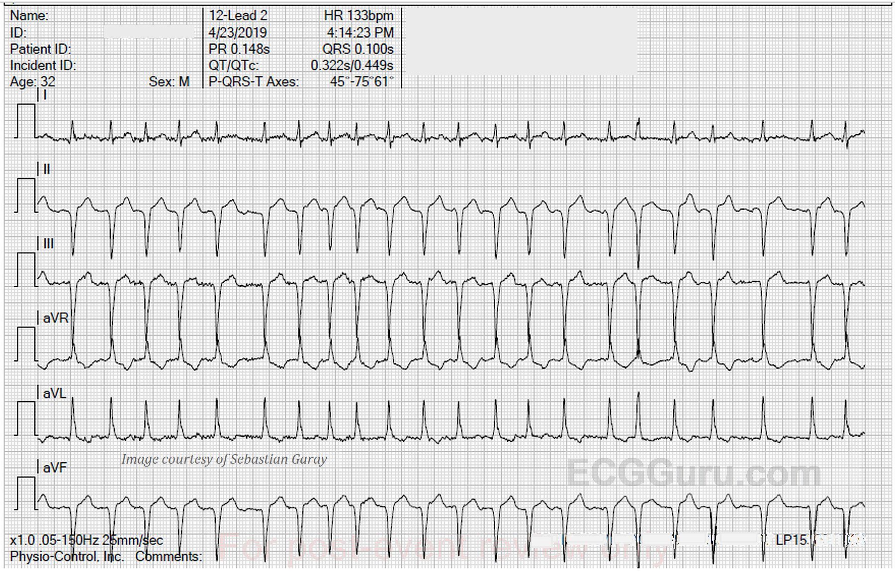
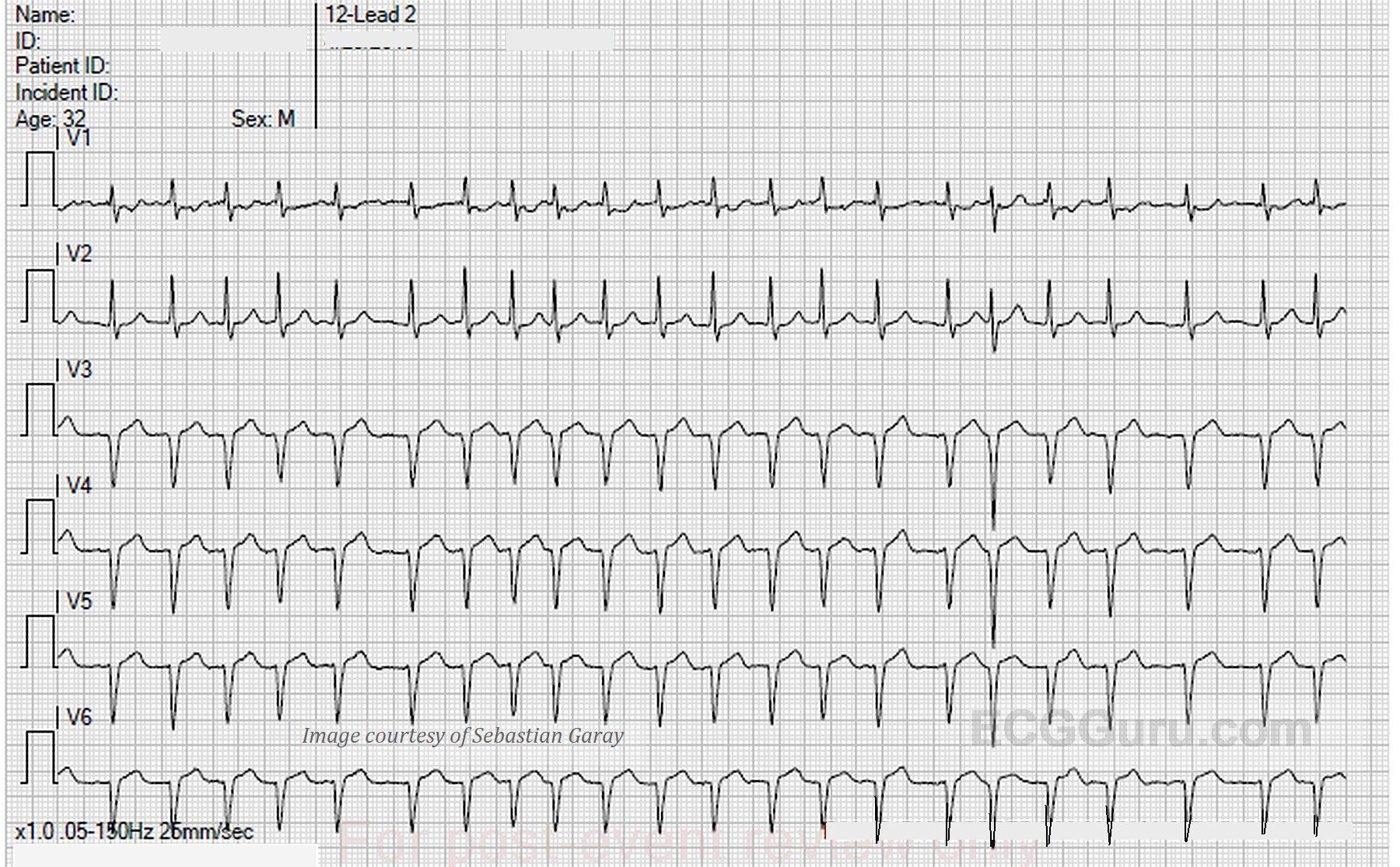
The ECG:      We are able in this case to provide a 12-lead ECG with each lead recorded for the entire width of the paper. This has the advantage of producing twelve ten-second rhythm strips.  Page one contains the limb leads, and page two shows us the precordial leads.

The rhythm is atrial fibrillation, with a heart rate of 133 bpm and an irregularly irregular rhythm. The QRS axis is extreme left at about -75 degrees.  This has caused Leads II, III, and aVF to be negatively deflected, and aVR and aVL to be positive. Lead I is biphasic, low voltage, and mostly positive, indicating that the axis travels almost perpendicular to Lead I, but slightly toward it.

The machine mistakenly gives us a reading for PR interval and P wave axis, even though there are no P waves.  The QRS is on the wide side without being abnormal at .10 seconds (100 ms). The QTc is within normal limits, although it might be considered “borderline”, with 431-450 usually considered borderline.

All our content is FREE & COPYRIGHT FREE for non-commercial use

Please be courteous and leave any watermark or author attribution on content you reproduce.