Displaying 1 - 10 of 18
Dawn's picture

Inferior Wall M.I. In A Patient With Left Bundle Branch Block

The Patient:    A 64-year-old man complaining of chest pain and shortness of breath for 20 minutes.  Long-standing history of triple vessel disease, severe aortic stenosis, hypertension, thrombocytopenia.  Meds unknown.  He was not considered to be a candidate for valve surgery.


The ECG: There is normal sinus rhythm with a rate of 90 bpm.  P waves are not visualized well in all leads, so remember that the three channels of this ECG are run simultaneously.  If you see a P wave in Leads I and II, they are also present in Lead III.  The PR interval is WNL.


The QRS complexes are wide, at .122 seconds (122 ms).  The criteria for left bundle branch block are met. (Supraventricular rhythm, wide QRS, upright QRS in Leads I and V6, negative QRS in V1).  The frontal plane axis is within normal limits, but toward the right, at 87 degrees.  The QRS complexes transition at V4 from negative to positive, but Leads V1 – V3 have no initial r waves.  These are possibly pathological Q waves, likely from a past anterior-septal M.I.


Dawn's picture

Rate-related Left Bundle Branch Block

The patient:  This ECG is from an 87-year-old man who was transported to the Emergency Department by paramedics. His chief complaint, as reported by caregivers, was lethargy, fever, and a declining mental status.  He appeared tired and slightly confused, and was normotensive.

The ECG:  There are a rhythm strip with two leads, II and III, and also a standard 12-lead ECG. The RHYTHM STRIP shows a tachycardiac rhythm that slows very slightly toward the end.  The rate is around 107 bpm, with an R to R interval of approximately 543 ms in the earlier, regular portion.  There are regular P waves present, all followed by QRS complexes.  Most of the QRS complexes are normal width, but the 2nd, 5th, and 8th are slightly wide at 130 ms, or .13 seconds.

These wider QRS complexes represent aberrant conduction with LBBB occurring intermittently.  Aberrant conduction often occurs due to a faster heart rate, but the only clue here is the intermittent conduction disturbance seems to disappear when the rate slows very slightly.  It is hard to determine mechanism of aberrant conduction when we have only a ten-second rhythm strip.

The 12-LEAD ECG  has essentially the same rate and rhythm, except all the beats in the first ¾ of the ECG are conducted aberrantly, in a LEFT BUNDLE BRANCH BLOCK pattern.  This indicates that the LBB is refractory at this time.  Beat No. 15 is premature (PAC).  The pause after the PAC allows the left bundle branch to repolarize, conducting one single beat normally.

Dawn's picture

Left Bundle Branch Block

This ECG is taken from an elderly man with heart failure. 

The ECG   The first feature that might capture your attention is the wider-than-normal QRS complex, which is 160 ms (.16 seconds).  The rate is 58 bpm. We do not know the patient’s medications or baseline rate.  There are P waves present, and so the rhythm is SINUS BRADYCARDIA. The P waves are broad , > 110 ms in Lead II (red lines in close up) and bifid, with greater than 40 ms between the two peaks in Lead II (blue lines).  In V1, the P waves are biphasic, with the terminal negative portion greater than 40 ms duration (red lines). This meets the ECG criteria for LEFT ATRIAL ENLARGEMENT, or preferably, LEFT ATRIAL ABNORMALITY. (https://LITFL.com/left-atrial-enlargement-ecg-library/) ECG criteria are not highly accurate for detecting atrial enlargement, and abnormal findings should be confirmed by anatomic measurement. (https://www.ncbi.nlm.nih.gov/pmc/articles/PMC2244611/).

The QRS complexes, as mentioned, are wide. Because there is sinus rhythm, we know the delay in conduction is due to interventricular conduction delay, and not to ventricular rhythm.  This ECG meets the criteria for LEFT BUNDLE BRANCH BLOCK. 

·        Supraventricular rhythm 

·        Wide QRS (>.12 seconds)

Dawn's picture

Wide Complex Tachycardia

The Patient   A 64-year-old woman has called 911 because she has chest discomfort radiating to her left arm, palpitations, weakness, and a headache.  She had a valve replacement (we do not know which valve) two weeks ago and has a healing incision over her sternum.  She is found sitting in a chair, pale, cool, and diaphoretic. Her blood pressure is 94/palp.  Her pulse rate is 196 bpm and weak. She is afebrile.

ECG #1   This ECG shows a wide-complex tachycardia at 196 bpm.  The QRS complexes are .132 seconds in duration, per the ECG machine. The rate is too fast to appreciate whether there are P waves present.  We did not see the onset of the tachycardia, but with a rate this fast and regular, it is most likely a reentrant rhythm, rather than sinus tachycardia.  An abrupt onset of the rhythm would point to a diagnosis of a reentrant rhythm, either ventricular tachycardia (VT) or paroxysmal supraventricular tachycardia (PSVT). 

There is an important rule in emergency medical care:  a wide-complex tachycardia should be treated as VT until and unless it is proven to be something else.  The most likely alternate interpretation is PSVT with aberrant conduction, which usually takes the form of left or right bundle branch block. Fortunately, the paramedics on this call have a protocol for treating WCT that includes electrical cardioversion for the unstable patient, and amiodarone for the stable patient.  This protocol serves both possibilities, VT and PSVT, well.  The patient’s perfusion status and BP made her borderline in this determination, but she was alert and oriented, so the paramedics opted for administering the amiodarone while they prepared to electrically cardiovert.

Dawn's picture

Wide Complex Tachycardia

The Patient:   The details of this patient’s complaints and presentation are lost, but we know he was a 66-year-old man who was being treated in the Emergency Department. His rhythm went from sinus tachycardia with non-respiratory sinus arrhythmia to multi-focal atrial tachycardia (MAT) to wide-complex tachycardia. The WCT lasted a few minutes and spontaneously converted to an irregular sinus rhythm.

Wide-complex tachycardia:  Ventricular tachycardia or aberrantly-conducted supraventricular tachycardia?  When confronted with a wide-complex tachycardia, it can be very difficult to determine whether the rhythm is ventricular or supraventricular with aberrant conduction, such as bundle branch block. The patient’s history and presentation may offer clues.  It is very important, if the patient’s hemodynamic status is at all compromised (they are “symptomatic”), the WCT should be treated as VENTRICULAR TACHYCARDIA until proven otherwise.  

There have been many lists made of the ECG features that favor a diagnosis of ventricular tachycardia. Here are two such lists:  Life In The Fast Lane, and National Institute of Health.

The ECG:  This ECG shows a regular, fast, wide-QRS rhythm.  The rate is 233 bpm.  It had a sudden onset and sudden offset (not shown on this ECG), and the rhythm lasted about 3-5 minutes. The patient felt the change in rate, but did not become hypotensive or unstable.  Some features that relate directly to the most commonly-referenced VT vs. SVT charts are:

Dawn's picture

Wide QRS Complex With First-degree AV Block

The Patient:  This ECG was taken from a 73-year-old man with a history of heart failure with preserved ejection fraction, severe left ventricular hypertrophy, Type II diabetes, and stage 4 chronic kidney disease.  He also suffered deep vein thrombosis and is on anticoagulation.  He has a recent diagnosis of IgA myeloma.  He presented with a complaint of nausea and vomiting and was found to have a worsening of acute kidney infection.  There was suspicion of renal and cardiac amyloidosis, but the patient refused biopsy to confirm this.  He was started on chemotherapy for multiple myeloma and will be followed as an outpatient.

The ECG:  The rhythm is sinus at around 60 bpm, although the rate varies a little at the beginning of the strip.  The QRS complex is wide at .12 seconds, or 120 ms., representing interventricular conduction delay (IVCD).  The PR interval is .32 seconds, or 320 ms. This constitutes first-degree AV block.  There is left axis deviation in the frontal plane and poor R wave progression in the horizontal plane.

Dawn's picture

Left Bundle Branch Block With Second-Degree AV Block, Type II

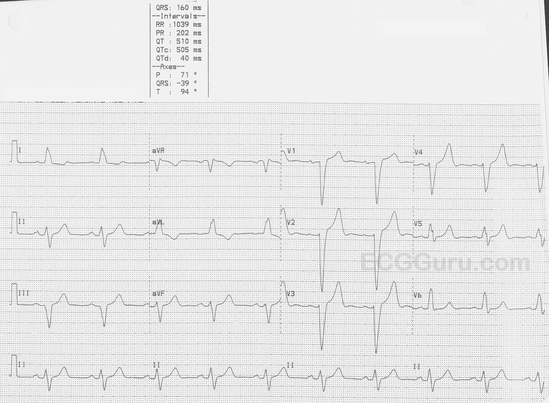
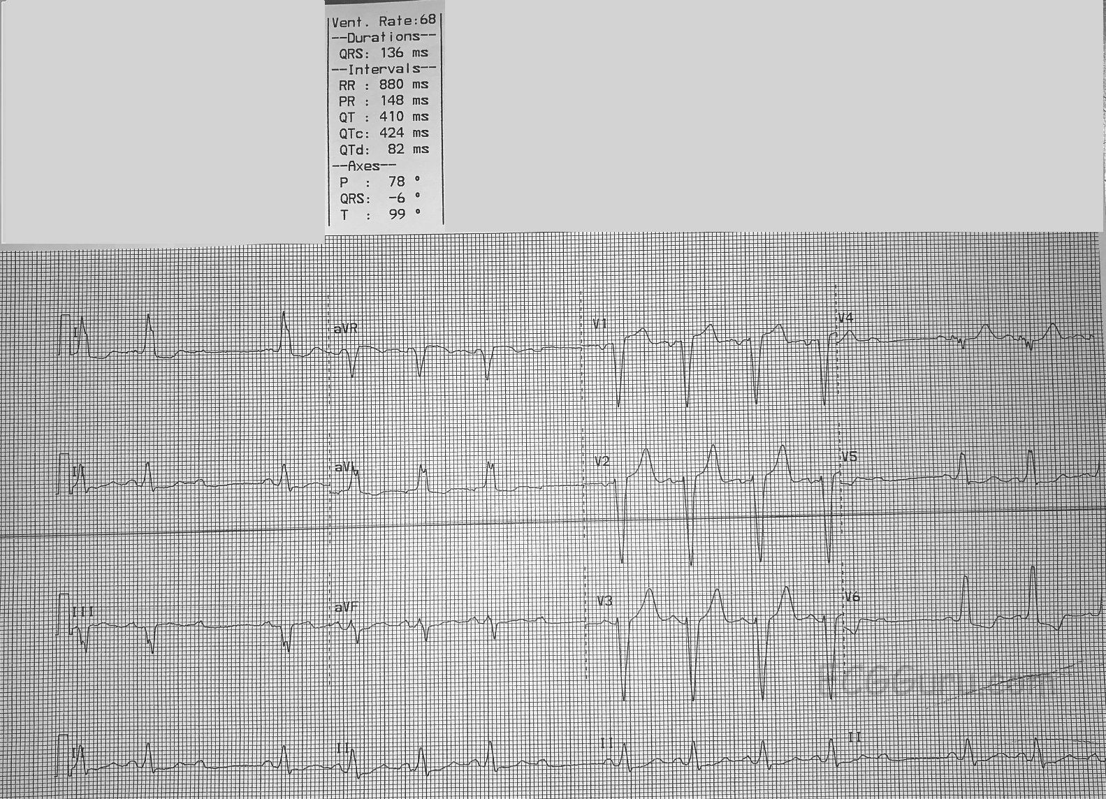
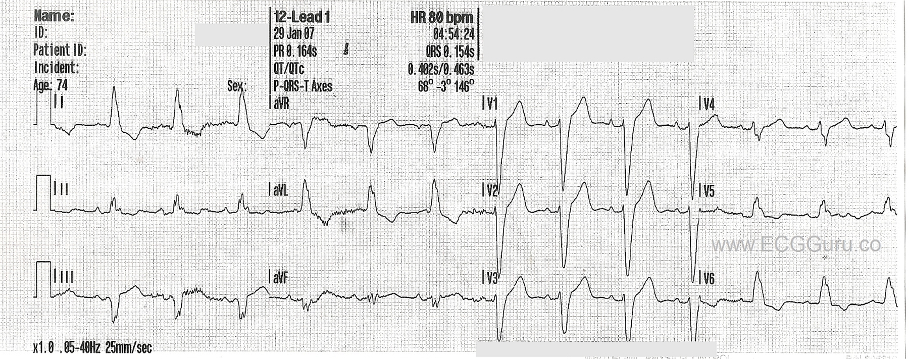
 This ECG was obtained from an 84-year-old woman who was scheduled for surgery.  When the anesthesiologist did this ECG, the surgery was cancelled. It is a very good example of fascicular-level blocks. 

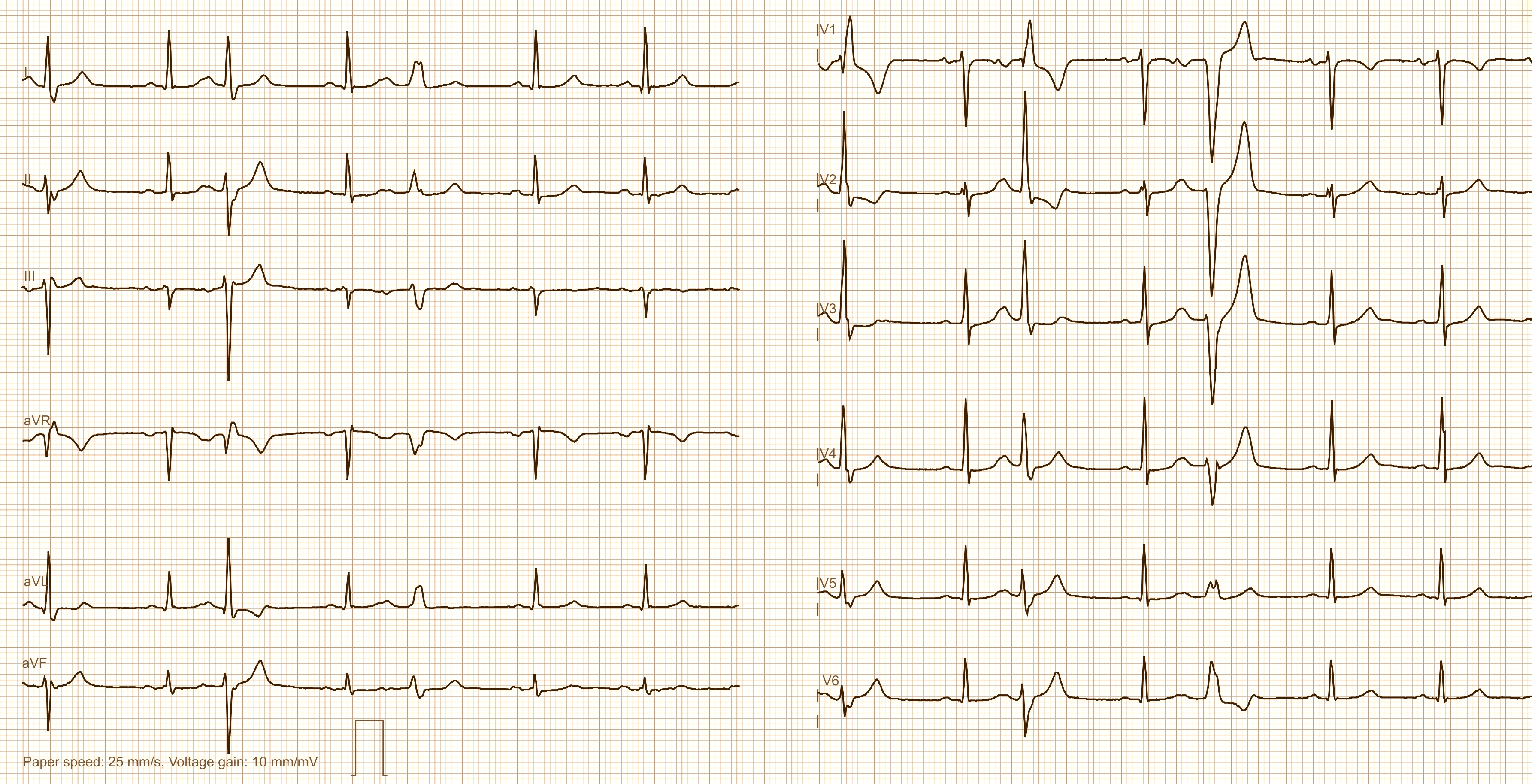
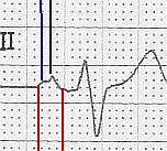
The underlying rhythm is a regular sinus rhythm at about 95 bpm.  There are some non-conducted P waves which are part of the sinus rhythm (not premature beats).  When the P waves DO conduct, the PR interval is steady at about .15 seconds (148 ms).

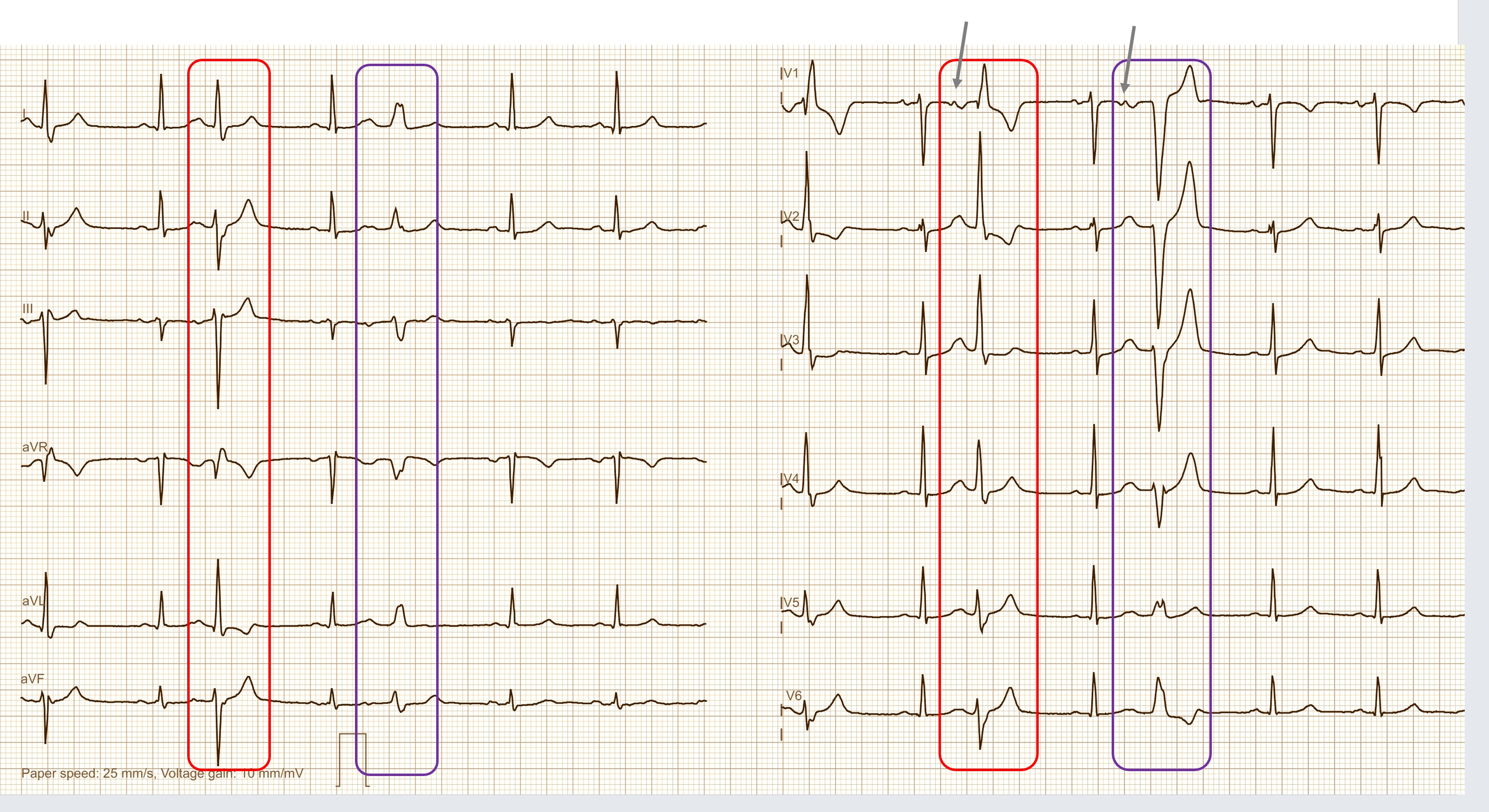
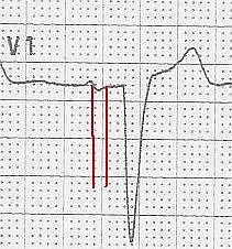
In addition, there is a LEFT BUNDLE BRANCH BLOCK.  The ECG criteria for LBBB are:  1) A supraventricular rhythm, 2) A wide QRS, and 3) A negative QRS in Lead V1 and a positive QRS in Leads I and V6.  The QRS duration in this ECG is 136 ms.

There are generally two fascicles (branches) in the left bundle branch, and one main fascicle in the right bundle branch.  So, a LBBB represents a “bi-fascicular block”.  That means that A-V conduction is proceeding down only one fascicle (the right bundle branch).  In that fascicle, there is an “intermittent” block.  When the RBB is not blocked, we see a QRS.  When it is blocked, we see none.  This is then termed an “intermittent tri-fascicular block” – otherwise known as SECOND-DEGREE AV BLOCK, TYPE II.  Type II blocks nearly always have a wide QRS due to the underlying bundle branch pathology.  You may see RBBB, LBBB, or RBBB with left anterior fascicular block (hemiblock).  Very rarely, the combination might include left posterior hemiblock.  The intermittent block in the “healthiest” fascicle(s) is what makes this a second-degree block, and not a complete heart block (third-degree AVB).

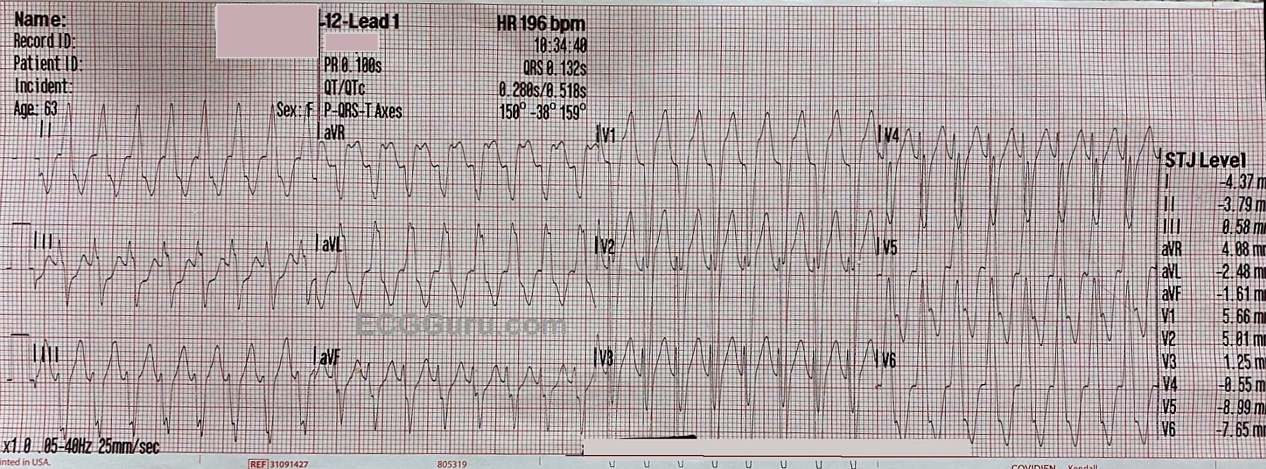
The clinical implications of this block are that the heart is operating on only one fascicle, and that fascicle is showing obvious signs of distress.  A third-degree AVB could be imminent.  In addition, LBBB causes a wide QRS, which decreases cardiac output.  Second-degree, Type II AVBs can result in very slow rates, and sometimes cause more hemodynamic instability that some third-degree AV blocks.

This patient was scheduled for pacemaker implantation instead of the originally-scheduled surgery. 

Dawn's picture

Left Bundle Branch Block

This ECG shows a “classic” left bundle branch block pattern. 

The ECG criteria for left bundle branch block are:

·        Wide QRS (.12 seconds or greater)

·        Supraventricular rhythm (ventricular rhythms do not travel via the LBB)

·        The QRS in V1 is negative, and the QRS in Leads I and V6 are positive. 

The left bundle branch (LBB) can be blocked permanently, temporarily, intermittently, or in the because of a fast rate.  When the LBB is blocked, conduction proceeds from the AV junction down the right bundle branch, depolarizing the right ventricle.  The impulse travels from the right ventricle across the left ventricle, cell by cell.  Conduction is slower this way, and there is asynchrony of the ventricles. This slow conduction and asynchrony of the two ventricles causes widening of the QRS complex.

NOTE:  It is "normal" for wide-complex rhythms to have ST segment elevation in leads with negative QRS complexes and ST depression in leads with positive QRS complexes.  This can make it a bit difficult to determine the ST changes of acute M.I. 


Dawn's picture

Left Bundle Branch Block and Artifact

This ECG offers several teaching opportunities.  First, it is an example of left bundle branch block (LBBB).  It was obtained from a 53-year-old man who was undergoing a cardiac cath for chest pain.  Unfortunately, we do not have access to his past medical history or the results of his cath.  The ECG criteria for a diagnosis of LBBB are:  1) wide QRS complex; 2) supraventricular rhythm; 3) negative QRS in V1 and positive QRS in V6 and Lead I.  This ECG shows normal sinus rhythm at a rate of 88 bpm and a wide QRS at 158 ms (.158 seconds).  The QRS in V1 is negatively deflected and in V6 and Lead I it is positive.

In LBBB, as with any condition that significantly widens the QRS, there will be ST-T changes.  The ST segment will deviate in the opposite direction of the QRS.  In other words, there will be ST elevation in leads with negative QRS complexes and ST depression in leads with positive QRS complexes.  LBBB causes significant difficulty for those trying to diagnose acute ST elevation using ECG alone.  Excessive ST elevation in a lead where elevation is expected OR ST elevation in a lead where depression is expected should be considered to be abnormal.  At this point, you may find it useful to review Sgarbossa's Criteria regarding determining the presence of acute M.I. in the presence of LBBB.

LBBB can be a serious functonal problem for the patient, as the slow ventricular conduction that causes the wide QRS results in less-than-optimal cardiac output.  This associates LBBB with congestive heart failure, both as a cause of CHF and a result of CHF.  Many people with LBBB and CHF can be helped by cardiac resynchronization therapy - pacing both ventricles synchronously to narrow the QRS and improve cardiac output.  For an excellent article on cardiac pacing in general and CRT (page 2299), go to the 2013 European Society of Cardiology Guidelines as reported by the European Heart Journal, (2013) 34, 2281–2329 doi:10.1093/eurheartj/eht150

Dawn's picture

Left Bundle Branch Block

This is a good example of sinus rhythm with left bundle branch block.  There is some irregularity due to a PAC at the beginning.  The QRS is wide at 144 ms (.14 seconds).  There is also first-degree AV block, with a prolonged PR interval of 228 ms.  The criteria for diagnosis of left BBB are:  wide QRS, supraventricular rhythm, and a negatively-deflected QRS in V1 with a positive QRS in Leads I and V6.  

Left bundle branch block can be associated with many forms of heart disease, including CHF.  It can be permanent, transient, intermittent, or rate-related.  The wide QRS of LBBB significantly decreases cardiac output, causing poor perfusion symptoms in some people.

This ECG is a good one for your students who are just transitioning from reading rhythm strips to reading 12-lead ECGs.  It shows the value of multi-lead assessment of rhythms. You will notice that P waves are difficult to see in some leads.  Armed with the knowledge that the four channels on this ECG are run simultaneously, you can show the students how finding P waves in one lead will allow you to find them in the leads that are above and below that lead. 

Similarly, it can be difficult to see the QRS width in some leads.  The leads in the same vertical column can help you see the QRS's true width, even if part of the QRS is "flat" in the isoelectric baseline.

Pages

All our content is FREE & COPYRIGHT FREE for non-commercial use

Please be courteous and leave any watermark or author attribution on content you reproduce.