Displaying 1 - 10 of 10
Dr A Röschl's picture

AVNRT TYPICAL FORM

18-year-old male; palpitations lasting hours, beginning at the age of 10. What arrhythmia is present? Let's first consider the heart rate: with a heart rate of 194 beats/min, the heart rate is too low for atrial flutter (1:1) (except in patients who have been pre-treated with medication), and the rate would be unusually high for atrial flutter with 2:1 conduction. Due to the regularity of the heart rhythm, atrial fibrillation can also be ruled out. This leaves atrial tachycardia, junctional tachycardia, AVNRT, and AVRT as possibilities.

Dr A Röschl's picture

VT or SVT? Here, Both!

Complex ECGs like this one have to be approached systematically. Firstly, we can see a normal sinus rhythm. A is the first beat of a wide complex tachycardia. This must be a ventricular tachycardia. Although there is a P-wave before the first beat of the tachycardia, it is not premature. Therefore, there is no SVT with aberrant conduction. The first beat of the tachycardia looks different from the subsequent beats because there is a fusion beat present. The VT conducts 1:1 back to the atria (a small negative P-wave can be seen at the end of the QRS complex in V1).

Dawn's picture

Teaching Series - Tachycardia and Left Anterior Fascicular Block

This series of three ECGs is from a 60-year-old man who was brought to the Emergency Department after being involved in a motor vehicle accident.  No injuries were found, but the patient was severely intoxicated by alcohol consumption.  He was conscious but agitated. 

ECG NO. 1     15:07:23

The first ECG was taken by fire-rescue personnel at the scene of the accident. His hemodynamic status was stable, and the rate was not addressed in the field. ECG No. 1 shows a supraventricular rhythm at 161 bpm, with a narrow QRS and P waves visible before each QRS. 

A notable feature of this ECG are the left axis deviation, by default diagnosed at left anterior hemiblock (left anterior fascicular block).  The .10 second QRS width is typical of LAHB, as is the rS pattern in Lead III.

Also  noted is the unusual R wave progression in the precordial leads.  The R waves are prominent in V2, and then fail to progress across the precordium, and the S waves persist. This is probably due to the hemiblock.  We do not know this patient’s medical history, except that he self-described as an “alcoholic”.  LAFB can be associated with coronary artery disease. 

ECG NO. 2      15:20:38

Now being evaluated in the Emergency Dept., we see the patient's heart rate is 163 bpm.  Some variability in the rate was noted with patient agitation and activity, so it was determined that the rhythm was probably sinus tachycardia.  There were no other significant changes in the ECG from the first one.  Unfortunately, we no longer have access to lab results, so we do not know his electrolyte or hydration status.  Labs confirmed ETOH intoxication. 

ECG NO. 3   15:43:26

Dawn's picture

Wide-complex Tachycardia: Ventricular Tachycardia

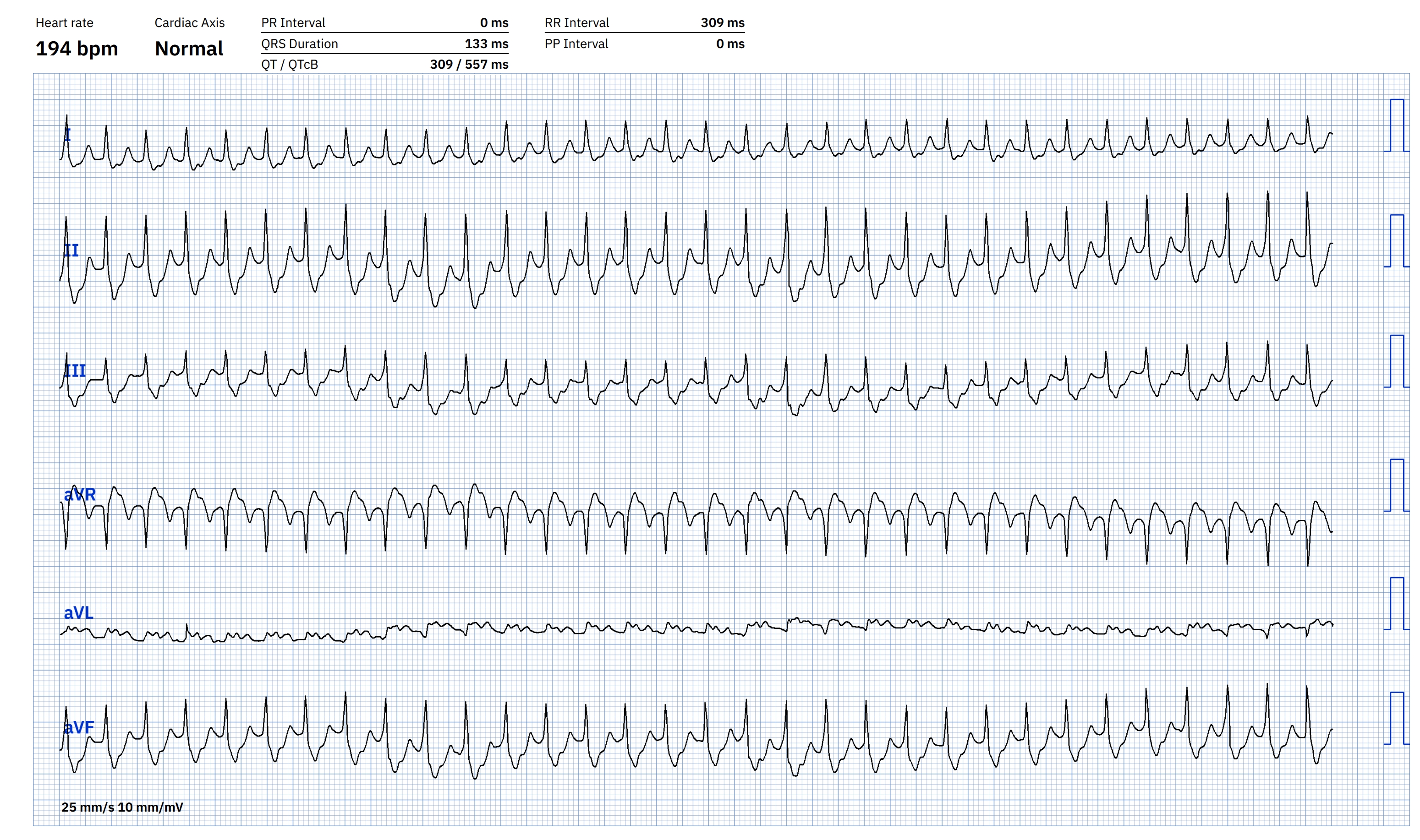
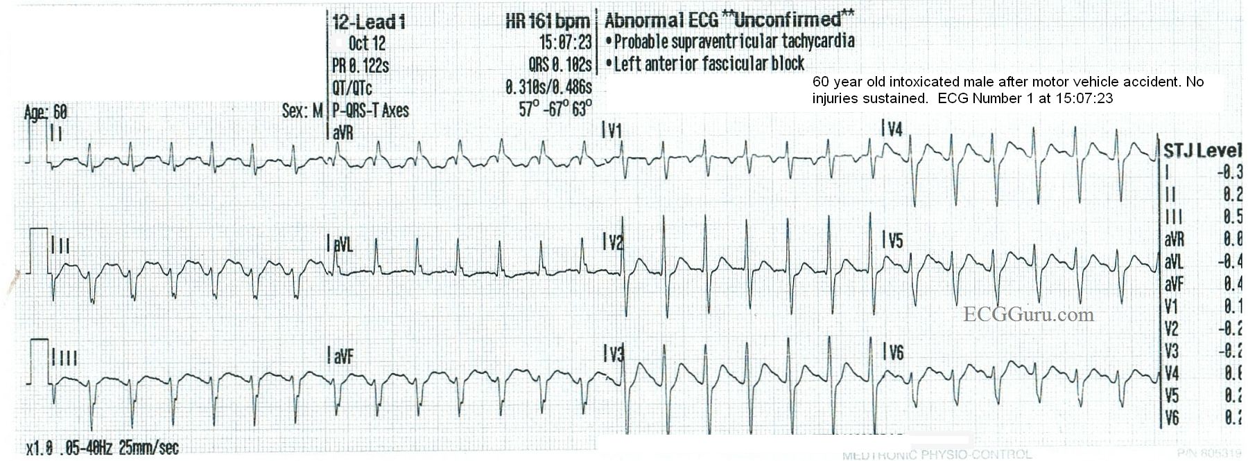
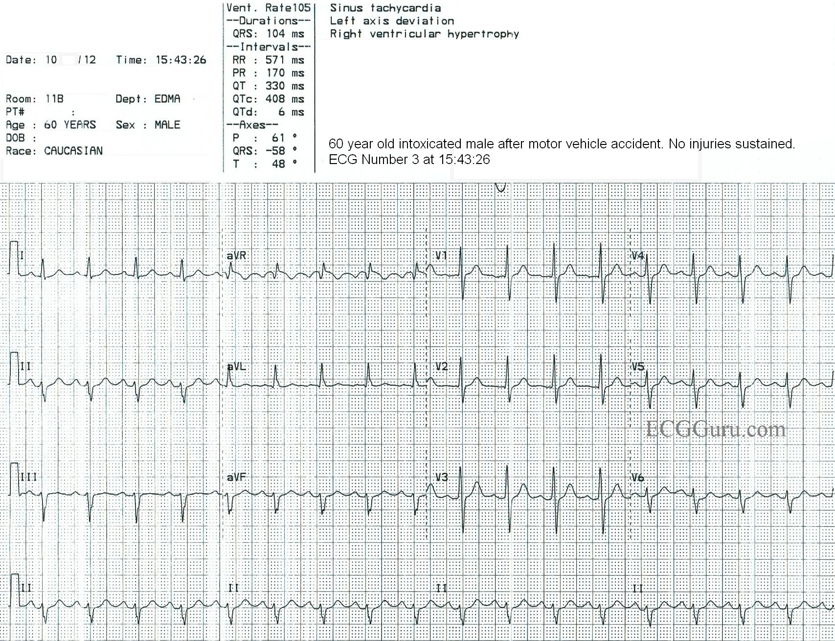
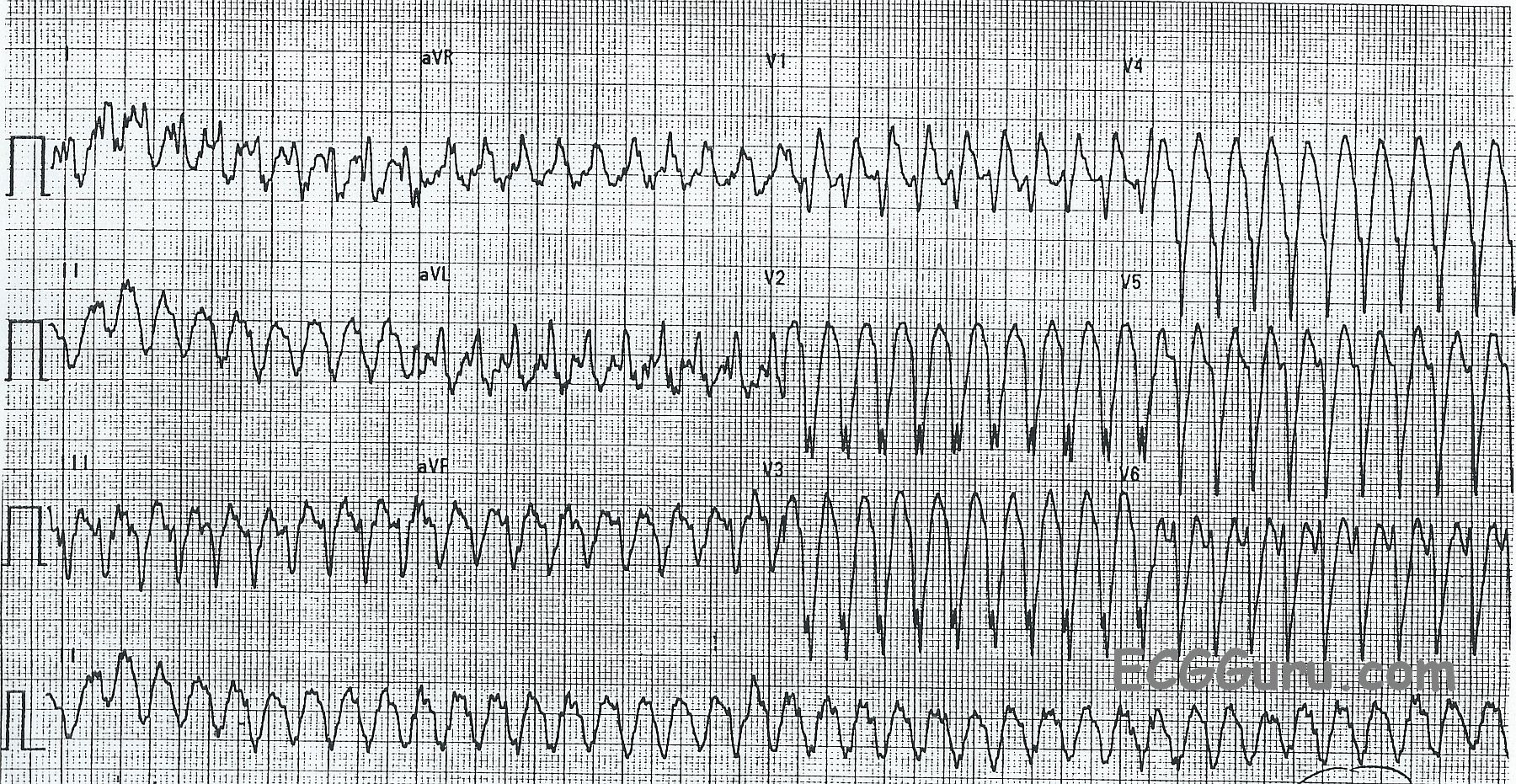
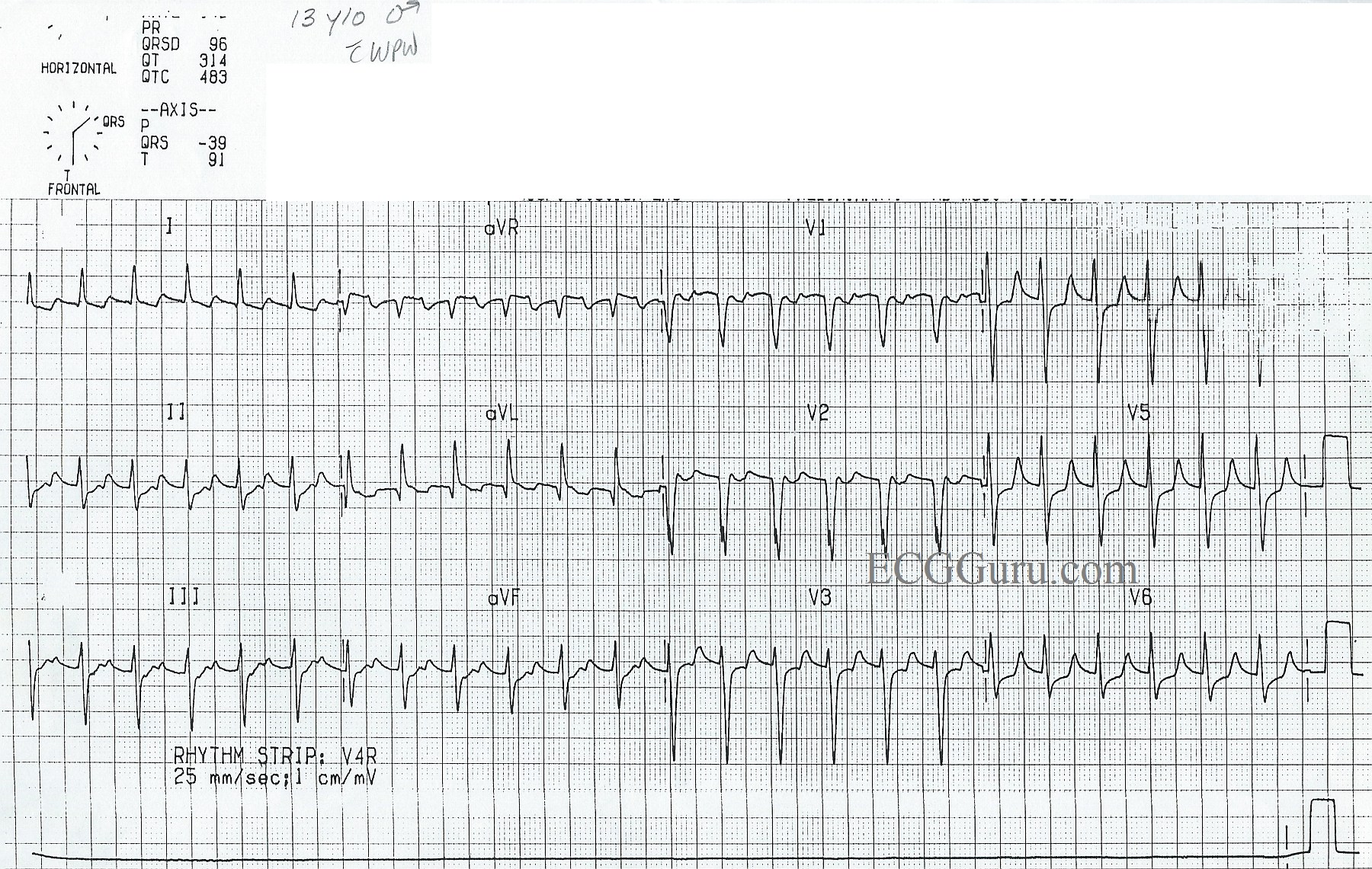
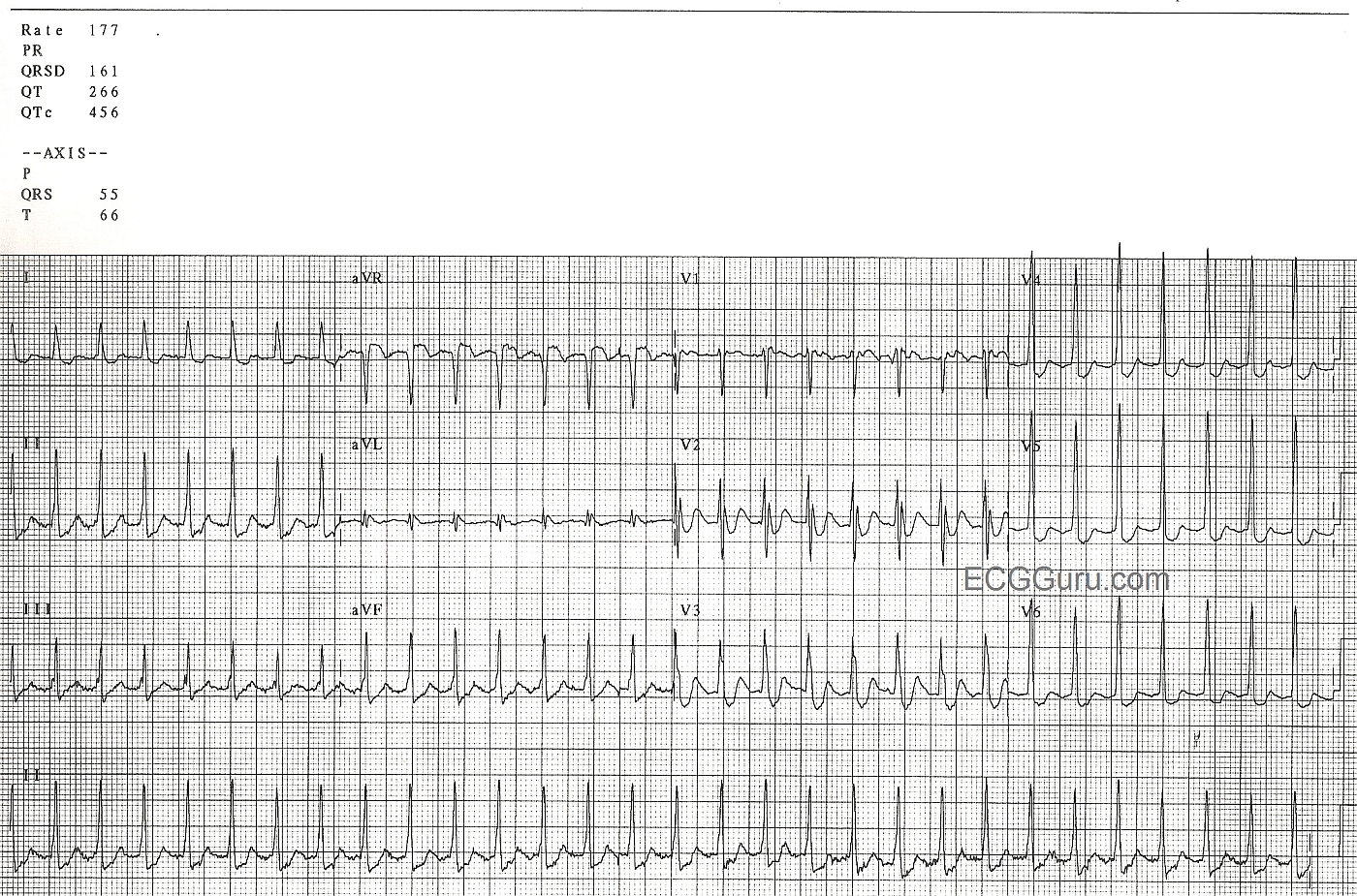
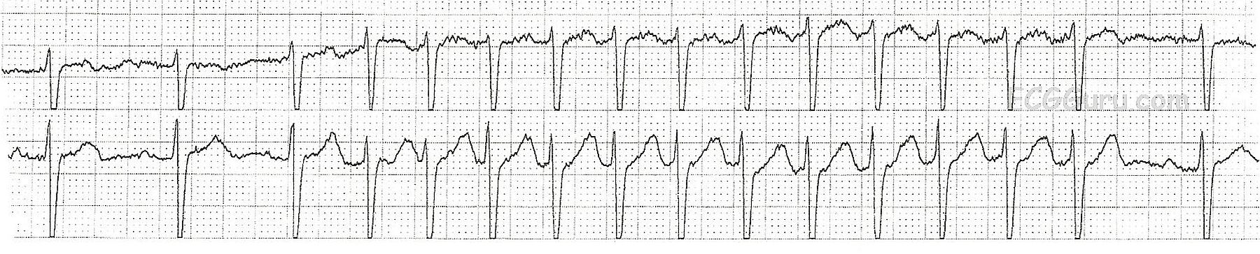
This ECG is from a man who was experiencing palpitations and light-headedness with near-syncope. On first look, you will see a wide-complex tachycardia (WTC) with a rate around 240 per minute.  It is difficult to assess for the presence of P waves because of the rate and the baseline artifact. 


The differential diagnosis of WCT includes ventricular tachycardia and supraventricular tachycardia with aberrant conduction, or interventricular conduction delay (IVCD). We should ALWAYS consider VENTRICULAR TACHYCARDIA first.  If the patient is an older adult with structural heart disease, WCT almost always proves to be VT. 

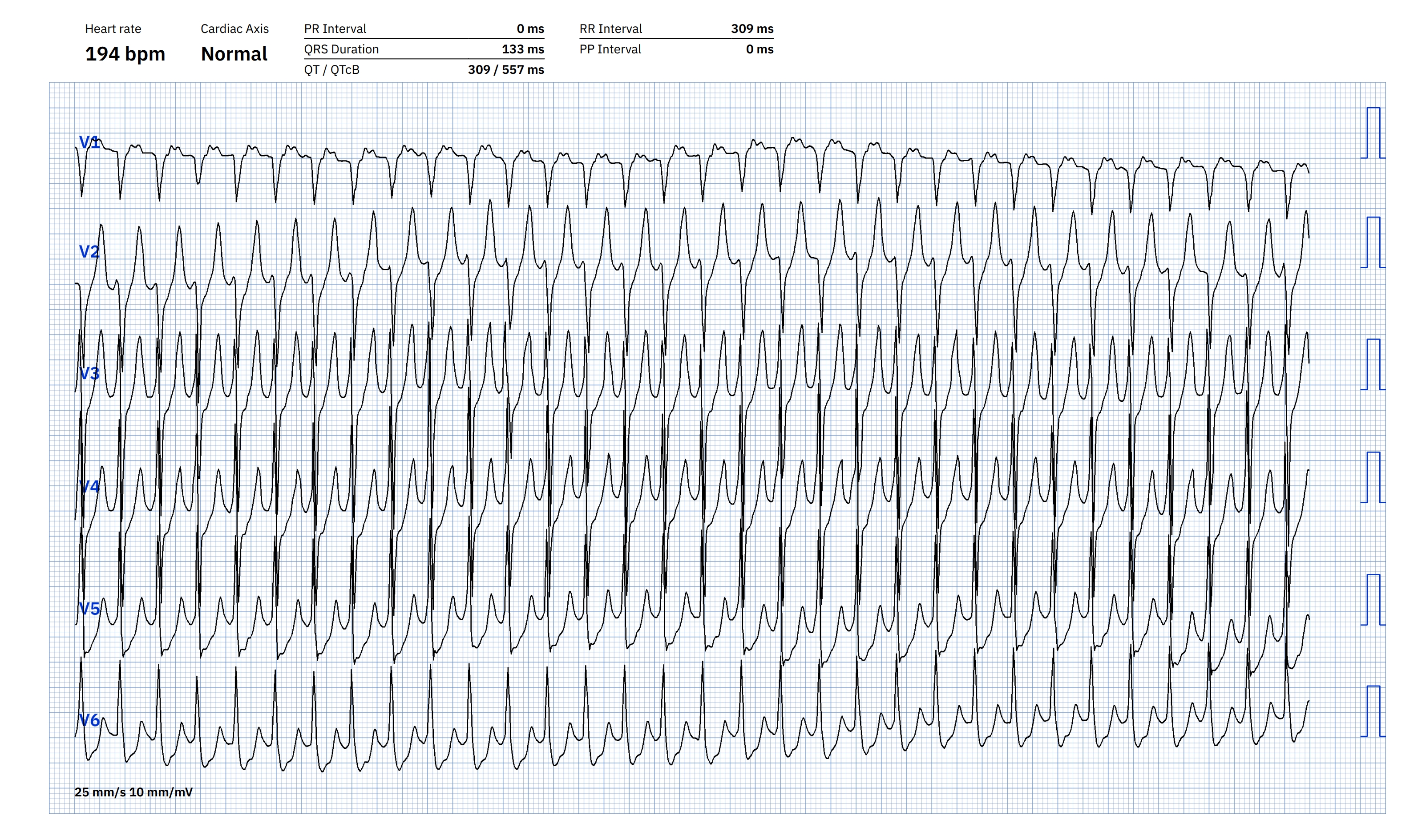
ABERRANT SVT?   In the setting of SVT with wide QRS, the most common aberrancy is right or left bundle branch block.  This ECG could be said to have a “RBBB” type pattern in V1, rSR’ and in Lead I and V6 with a wide S wave.  However, the other precordial leads do not have a RBBB pattern. 

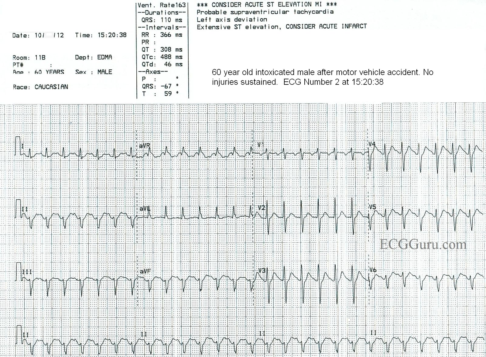
VENTRICULAR TACHYCARDIA? There are some features of this ECG that favor the diagnosis of VENTRICULAR TACHYCARDIA (VT).  They include, but are not limited to:

* Regular, wide QRS complexes, about .14 seconds in this ECG, but varies because of difficulty in measuring the beginning and end of the QRS in each lead.  The artifact obscures the exact points of beginning and ending. The QRS complexes, especially from V2 leftward, are very “ugly”, and don’t resemble patterns we would expect with bundle branch block.

* Horizontal plane axis extremely abnormal:  Leads II, III, and aVF are negative and aVR and aVL are positive.  The biphasic Lead I indicates a nearly vertical axis at around – 90 degrees.

* There is “almost” precordial concordance, but V1 is biphasic. 

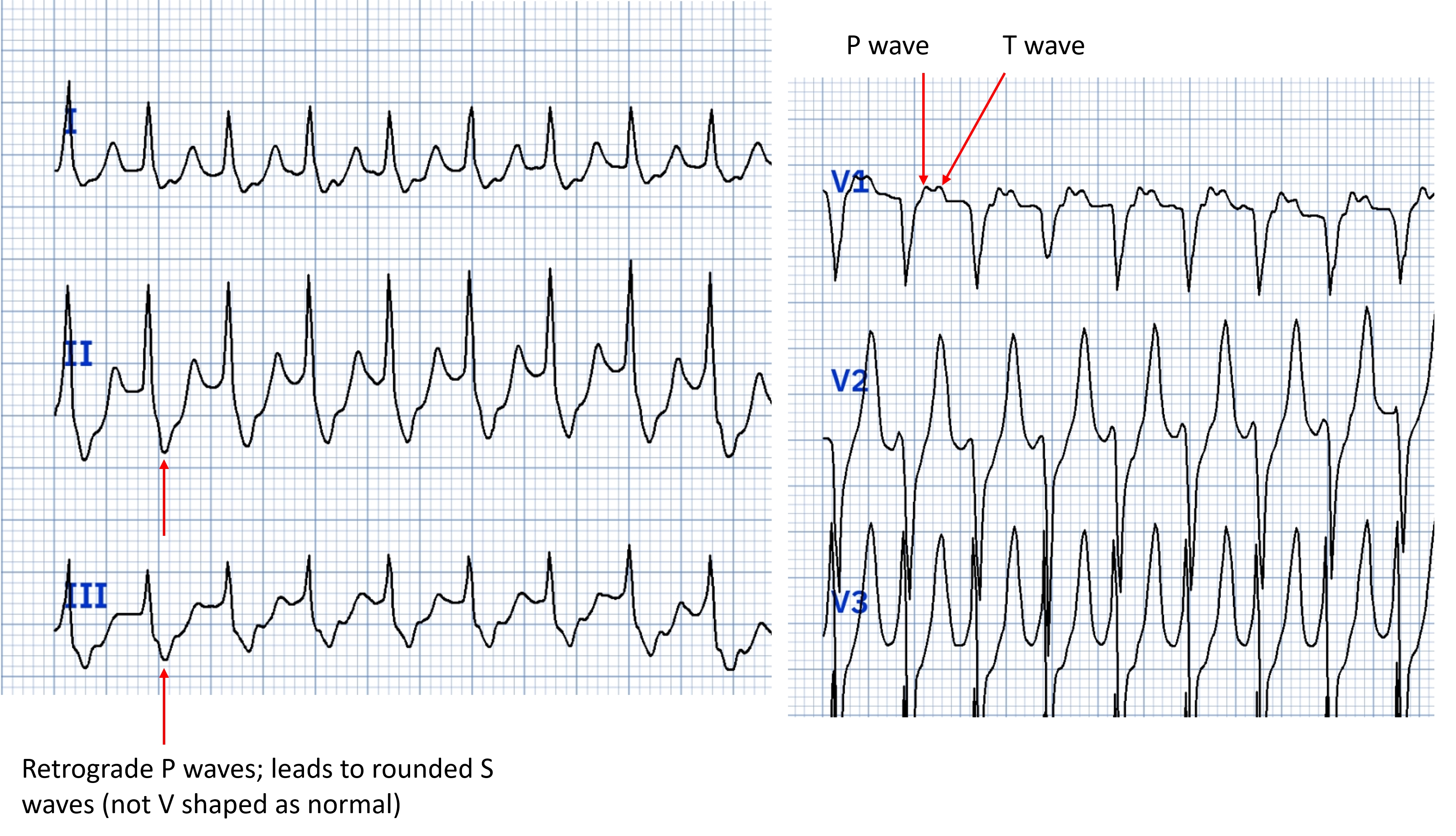
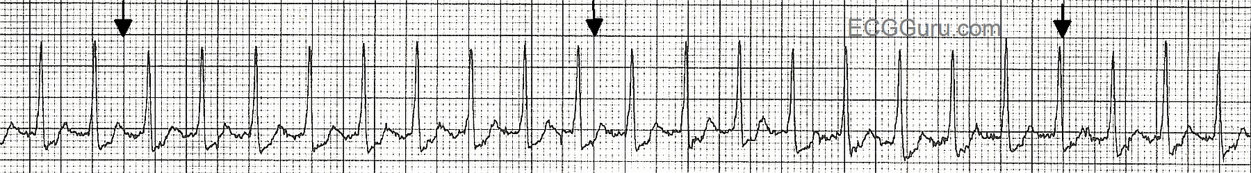
Unfortunately, we do not see capture beats or fusion beats, which would secure the diagnosis of VT. Disassociated P waves would also be a sure sign of VT, but the artifact in this ECG makes it impossible to say whether there are P waves. 

Dawn's picture

Supraventricular Tachycardia in Pediatric Patient With Wolff-Parkinson-White Syndrome

This ECG was obtained from a 13-year-old boy who had previously been in NSR.  At that time, he was diagnosed with Wolff-Parkinson-White syndrome, and he had delta waves.  He now presents with a supraventricular tachycardia.  People with W-P-W have one or more accessory pathways which allow atrial impulses to bypass the AV node and “pre-excite” the ventricles.  An accessory pathway, along with the normal pathway through the AV node, can form a functional circuit, allowing conduction to proceed down one pathway and return up the other, in a rapidly repeating circular motion.  This causes paroxysmal supraventricular tachycardia. 


In W-P-W, the most common type of SVT is AV reciprocating tachycardia (AVRT), also called AV reentry tachycardia.  Conduction through the accessory pathway can be forward (anterograde) or backward (retrograde).  The most common type of reentrant conduction in W-P-W is ORTHODROMIC, which we see here.  The impulse conducts forward through the AV node, and backward over the accessory pathway.  There is no delta wave, as there is no pre-excitation of the ventricles. 


Features of orthodromic AVRT are:

Fast rate (usually approximately 140-250 beats per minute)    

Narrow QRS complexes (unless BBB or IVCD present)   

P waves retrograde, if seen (negative in II, positive in aVR and V1)  

ST depression and T wave inversion common. 


Dawn's picture

Paroxysmal Supraventricular Tachycardia

This ECG is from a man in his 60's who is experiencing chest discomfort and palpitations.  The onset of the rapid heart rate and the symptoms was sudden, while he was at rest.  The rate did not slow when he was placed on oxygen, given IV fluids, and rested further. The rate is 177 / min.  

The rhythm is AV nodal reentry tachycardia (AVNRT), which is one of the rhythms that falls into the category of paroxysmal supraventricular tachycardia (PSVT).We can see signs of retrograde P waves in some leads (II, III, aVF, V1).  AVNRT is caused by a reentry circuit in the AV node.


Some instructors teach students that sinus tach is approximately 100-150 per minute, and atrial tach is usually 150-250 per minute.  If students only learn about differentiating these two rhythms by the rate difference, it will cause later problems.  Of course, there is actually an overlap in rates between the two rhythms.  For example, a febrile, dehydrated infant could easily reach this rate and be in sinus rhythm.  A young, healthy person on a treadmill could, too.  Clues to the ectopic origin of this rhythm are:  sudden onset (unfortunately, not witnessed here), regular rhythm with unwaivering rate, and the patient's situation (symptoms while at rest, no obvious reason for sinus tach).  Of course, we need to teach to the level of our students' abilities.  Consider whether they just memorizing rhythms criteria now, or are they learning about re-entry?

Dawn's picture

Supraventricular Tachycardia

This ECG shows AV nodal reentrant tachycardia in an elderly man.  Clinical information is not available.  AVNRT is the rhythm most often associated with the term, "supraventricular tachycardia".

Although we can't see the beginning of this rhythm, one of the identifying features of SVT is an abrupt (paroxysmal) onset.  In patients with AVNRT, there are two pathways in the AV node, a pathway with fast conduction and a long refractory period, and a pathway with slow conduction and a short refractory period.  Normal sinus impulses travel down the fast pathway and into the ventricles, but also start up the slow pathway in a retrograde direction. The retrograde impulse and the normal impulse traveling down the slow pathway collide, cancelling each other out.  If a PAC occurs, it will travel down the slow pathway while the fast pathway is still refractory.  By the time the impulse reaches the end of the slow pathway, it finds the fast pathway no longer refractory, and travels back up to the atria.  This forms a circular movement (circus movement) of the impulse, and it repeats itself rapidly until interrupted.  When each impulse reaches the ventricles, it travels into the interventricular conduction system and causes ventricular depolarization and contraction, usually at a rate of 140 - 250+.  Unlike sinus tachycardia, AVNRT does not adjust its rate according to the needs or activity of the patient.

There are many forms of supraventricular tachycardia, and they are not always easy to differentiate based on ECG criteria alone.  AVNRT of the type described above is the most common PSVT in structurally normal hearts.  For more information on supraventricular tachycardia, go to Life in the Fast Lane.  For a discussion on clinical management, we recommend Dr. Grauer's ECG Video 6 - Rhythm Mgmt-Part 3.

Dawn's picture

ECG Basics: Paroxysmal Supraventricular Tachycardia

This two-lead rhythm strip clearly shows the transition from normal sinus rhythm to a paroxysmal supraventricular rhythm.  In this case, the arrhythmia is AV nodal reentrant tachycardia, AVNRT.  The rate of the first rhythm, NSR, is around 75 per minute.  The fourth beat on the strip is a PAC which initiates the paroxysm of tachycardia lasting 12 beats.  The arrhythmia terminates spontaneously at that point.  The tachycardia rate is about 150/min.

The topic of supraventricular tachycardias can be a very complex one to teach.  For an excellent example of a concise lesson geared toward Primary Practice physicians, go to Dr. Grauer's VIDEO - Part III of his Arrhythmia series.

To cover the important points for the beginner-level student:

  *  It can be difficult to determine a rhythm is SVT if the rhythm is near 150 bpm and you DON'T see the beginning or end of the arrhythmia.  If the onset (or offset) is sudden, then this is not a sinus rhythm.  The sinus node speeds and slows more gradually - it doesn't change rates in one heartbeat.  This strip has an excellent view of BOTH the onset and the offset.

  *  The faster the rate, the more likely we are looking at a PSVT rather than sinus rhythm.  If a sinus tachycardia exists, we can almost ALWAYS see the reason for it in the patient's clinical situation.  We may see fever, dehydration, bleeding, fear, pain, exercise.  Therefore, a patient at rest with a rate of 150 would be suspect for PSVT.  A patient on a treadmill for 5 minutes would be considered to have a sinus rhythm.

  *  Any patient with a rate around 150 per minute should be evaluated for ATRIAL FLUTTER with 2:1 conduction.  Atrial flutter often conducts at that ratio, because a rate of 150 is fairly easy for the AV node to conduct, whereas the instrinsic rate of atrial flutter (250-350) is not.  A 12-lead ECG makes it easier to search for tell-tale flutter waves.

Dawn's picture

ECG Basics: Supraventricular Tachycardia

This strip is from a patient who experienced a sudden onset of palpitations and rapid pulse while at rest.  It shows a narrow-complex tachycardia, specifically a paroxysmal supraventricular tachycardia.  The subject of supraventricular tachycardias is a fascinating one, and is covered extensively throughout this website.  The mechanisms of SVT are many, and can be complex for the beginning student to understand.  Search the search terms on the left side of the page for entries from ASK the EXPERT and JASON's BLOG for more advanced information about SVT.

For the beginner, it is important to teach the difference between sinus tachycardia and "supraventricular tachycardia".  Of course, sinus tachycardia IS supraventricular - but current convention has us using the term "SVT" for atrial or junctional tachycardias, and especially for reentrant tachycardias.  Beginner students should understand the function of the sinus node, and it's ability to control the heart rate, based on direction given by the nervous system.  The sinus node increases and decreases the rate incrementally, or more gradually than the onset and offset of a reentrant tachycardia.  The appearance of a sudden onset of regular tachycardia following a PAC, producing a rhythm with a distinctly faster rate than the original sinus rhythm, is a sure sign of SVT.  When the onset or offset are caught on the rhythm strip, our job is SO much easier!

The heart rate helps with the diagnosis.  SVTs tend to be faster than sinus tachycardias.  SVTs tend to be faster than 150/min, while sinus rhythms TEND to be slower than 150.  And patients with sinus tachycardia usually have a readily determined reason for the tachycardia, such as fever, pain, fear, hypovolemia, hypoxia, or exertion.  So, a patient on a treadmill for an exercise stress test might very well have a heart rate over 150 / min.

When your students master the understanding of the different behaviors of the sinus node and the reentrant rhythms, and how important patient presentation is to the diagnosis, you will want to add atrial flutter and atrial fibrillation.  When these are mastered, be sure to remind your students that atrial flutter can conduct 2:1, and will mimic sinus tach.  A good rule of thumb is: Under 150/min.:  look for sinus tach first.  Around 150 / min.: look for atrial flutter with 2:1 conduction.  Over 150/min.: suspect PSVT.   


Dawn's picture

How do you teach the concept of reentry in PSVT to students with only a basic understanding of dysrhythmias?

Our expert today is Dr. Ken Grauer. He is a frequent contributer to the ECG Guru.


KEN GRAUER, MD is Professor Emeritus (Dept. Community Health/Family Medicine, College of Medicine, University of Florida in Gainesville).
Dr. Grauer has been a leading family physician educator for over 30 years. During that time he has published (as principal author) more than 10 books and numerous study aids on the topics of ECG interpretation, cardiac arrhythmias, and ACLS (including an ongoing Educational ECG Blog.

 
Answer:
PSVT is a reentry tachycardia. This arrhythmia carries many names, one of which is AVNRT = AV Nodal Reentry Tachycardia - recognizing that in the vast majority of cases, there is reentry occurring in or around the AV node. The AV node is not a homogeneous structure - instead functionally (and anatomically on a microscopic level) - there are 2 basic pathways. One of these conducts "fast" - and the other "slower". Conduction preferentially goes down the "fast" pathway (thereby 'blocking' and preventing conduction down the slow pathway). But if for any reason (like a PAC) the fast pathway is "blocked" - then conduction of the impulse will have to go down the "slow" pathway. If the timing is just right - conditions may be set up that allow "reentry" within the AV node - with the impulse going down the slow pathway and up the fast pathway. Less commonly, reentry within the AV node may be set up in which the impulse goes down the fast pathway and back up the slow pathway.


Think of the phenomenon of reentry as comparable to the situation when 50 young children are all holding hands and running around in a circle while holding hands. All it takes is for one disgruntled person to stick out their leg - and ALL 50 of the children who are holding hands will fall down. So it is with reentry - it is a circuit that is set up by fortuitous circumstances of conduction speed, refractory period duration, and usually a precipitating premature impulse. How do we treat AV Nodal Reentry Tachycardia (PSVT)? Either by vagal maneuvers or medication such as adenosine, diltiazem/verapamil, or beta-blockers - ALL of which at least transiently alter conduction properties within a portion of the AV node. Just like the disgruntled citizen who stuck out their leg and tripped up all 50 children - all it takes is brief alteration/interruption of the conduction circuit (by meds or vagal maneuver) to terminate an AV Nodal Reentry Tachycardia.


Simplistic illustration of the concept of reentry appears in the Figure below (excerpted from pp 567-574 of Grauer K, Cavallaro D: ACLS: Comprehensive Review [Vol 2] - 3rd Edition, Mosby Lifeline, St. Louis - 1993). For a more detailed look at reentry - visit: https://www.kg-ekgpress.com/reentry-svt/ - where you can download a pdf of the above 7 pages.


Ken Grauer, MD ([email protected])


Exerpted from pp 567-574 of Grauer  K, Cavallaro D: ACLS: Comprehensive Review (Vol. 2) - 3rd Edition, Mosby Lifeline, St. Louis - 1993.


All our content is FREE & COPYRIGHT FREE for non-commercial use

Please be courteous and leave any watermark or author attribution on content you reproduce.