Displaying 1 - 4 of 4
Dawn's picture

Left Bundle Branch Block

This ECG is taken from an elderly man with heart failure. 

The ECG   The first feature that might capture your attention is the wider-than-normal QRS complex, which is 160 ms (.16 seconds).  The rate is 58 bpm. We do not know the patient’s medications or baseline rate.  There are P waves present, and so the rhythm is SINUS BRADYCARDIA. The P waves are broad , > 110 ms in Lead II (red lines in close up) and bifid, with greater than 40 ms between the two peaks in Lead II (blue lines).  In V1, the P waves are biphasic, with the terminal negative portion greater than 40 ms duration (red lines). This meets the ECG criteria for LEFT ATRIAL ENLARGEMENT, or preferably, LEFT ATRIAL ABNORMALITY. (https://LITFL.com/left-atrial-enlargement-ecg-library/) ECG criteria are not highly accurate for detecting atrial enlargement, and abnormal findings should be confirmed by anatomic measurement. (https://www.ncbi.nlm.nih.gov/pmc/articles/PMC2244611/).

The QRS complexes, as mentioned, are wide. Because there is sinus rhythm, we know the delay in conduction is due to interventricular conduction delay, and not to ventricular rhythm.  This ECG meets the criteria for LEFT BUNDLE BRANCH BLOCK. 

·        Supraventricular rhythm 

·        Wide QRS (>.12 seconds)

Dawn's picture

Left Bundle Branch Block With Left Atrial Enlargement

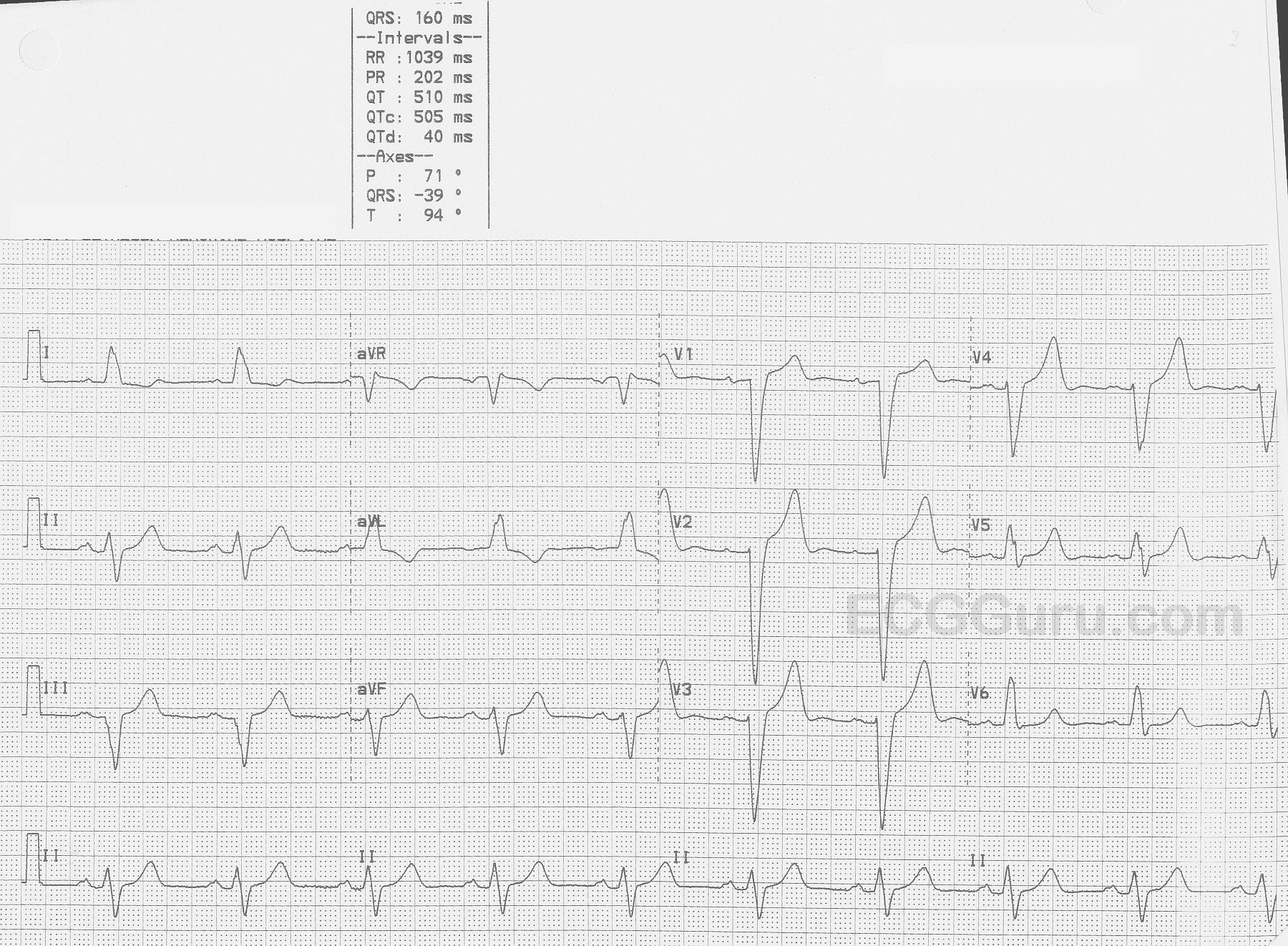
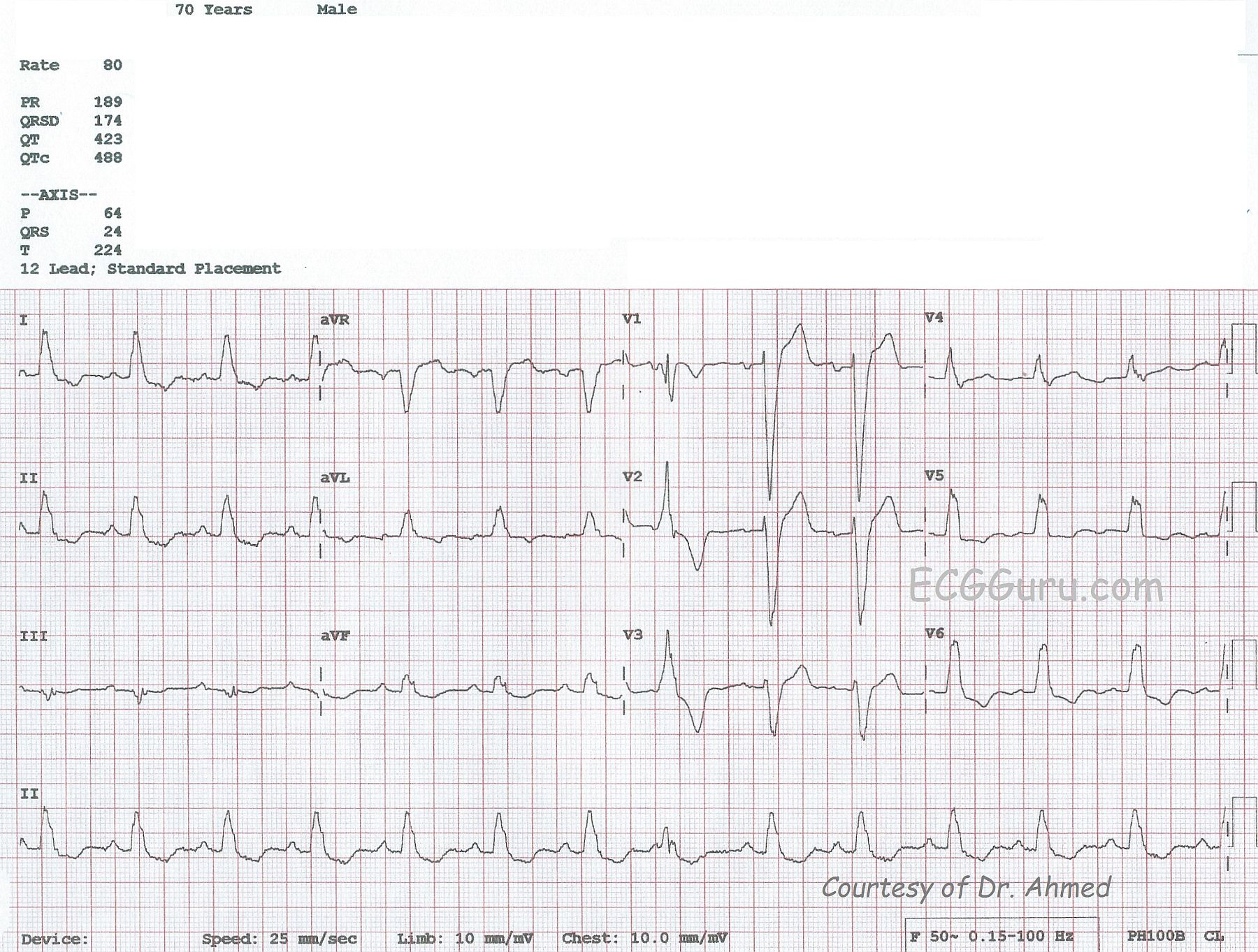
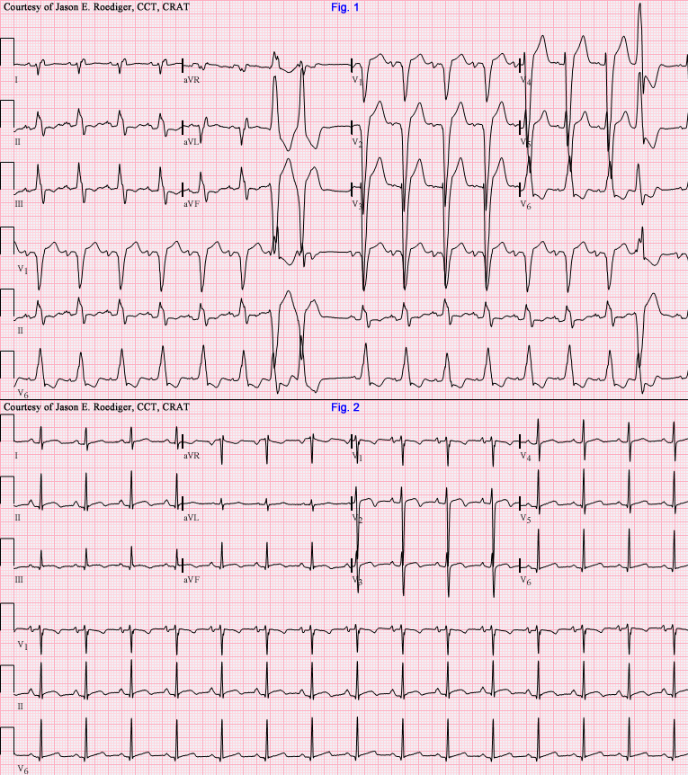
This ECG, kindly donated by Dr. Ahmed from India, is from a 70-year-old man shows a sinus rhythm at 80 bpm with left bundle branch block (LBBB), left atrial enlargement (LAE), and a premature ventricular contraction (PVC). The ECG criteria for LBBB is:  1) Wide QRS  - greater than or equal to .12 seconds;  2) Supraventricular rhythm;  3) QRS that is negative in V1 and positive in Leads I and V6. In leads with a positive QRS, we will see some ST depression, and in leads with a negative QRS, some ST elevation.  This is "normal" for the wide QRS rhythm, and does not indicate injury or ischemia, although it does not rule it out, either.  LBBB is an indicator of cardiac disease, but not specific to one etiology.

There is a PVC seen as the 8th beat from the left, and it gives you a chance to show your students a wide-complex beat that is NOT associated with a P wave and is premature, compared to the wide-complex SINUS beats with LBBB.  The PVC, being wide-complex, also has similar ST changes:  the ST segments and T waves are DISCORDANT with the QRS complexes.

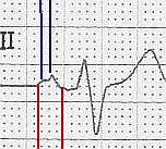
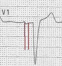
The P waves show some signs of enlargement of the left atrium.  The P waves in Lead II are tall and pointed, and the P waves in V1 are biphasic.  Left atrial enlargement in a patient with LBBB would not be surprising, as both are associated with left ventricular dysfunction.  Patients with these ECG patterns should be thoroughly evaluated for congestive heart failure.  Patients with LBBB, low ejection fractions, and heart failure are treated with cardiac resynchronization therapy, using a pacemaker that paces the atria and each ventricle, synchronizing both the A-V coupling interval and the depolarization of the ventricles for optimum cardiac output.

jer5150's picture

Jason's Blog: ECG Challenge of the Week for March 24th - 31st.

Patient's clinical data:  47-year-old black man.  Another serial ECG (seen below) showed a supraventricular tachycardia (SVT) at a rate of 164/min that was diagnosed by an Electrophysiologist as atrioventricular nodal reentrant tachycardia (AVNRT).  How does knowing the mechanism of AVNRT help you in determining the nature of the mechanism in this week's ECG.  

All our content is FREE & COPYRIGHT FREE for non-commercial use

Please be courteous and leave any watermark or author attribution on content you reproduce.