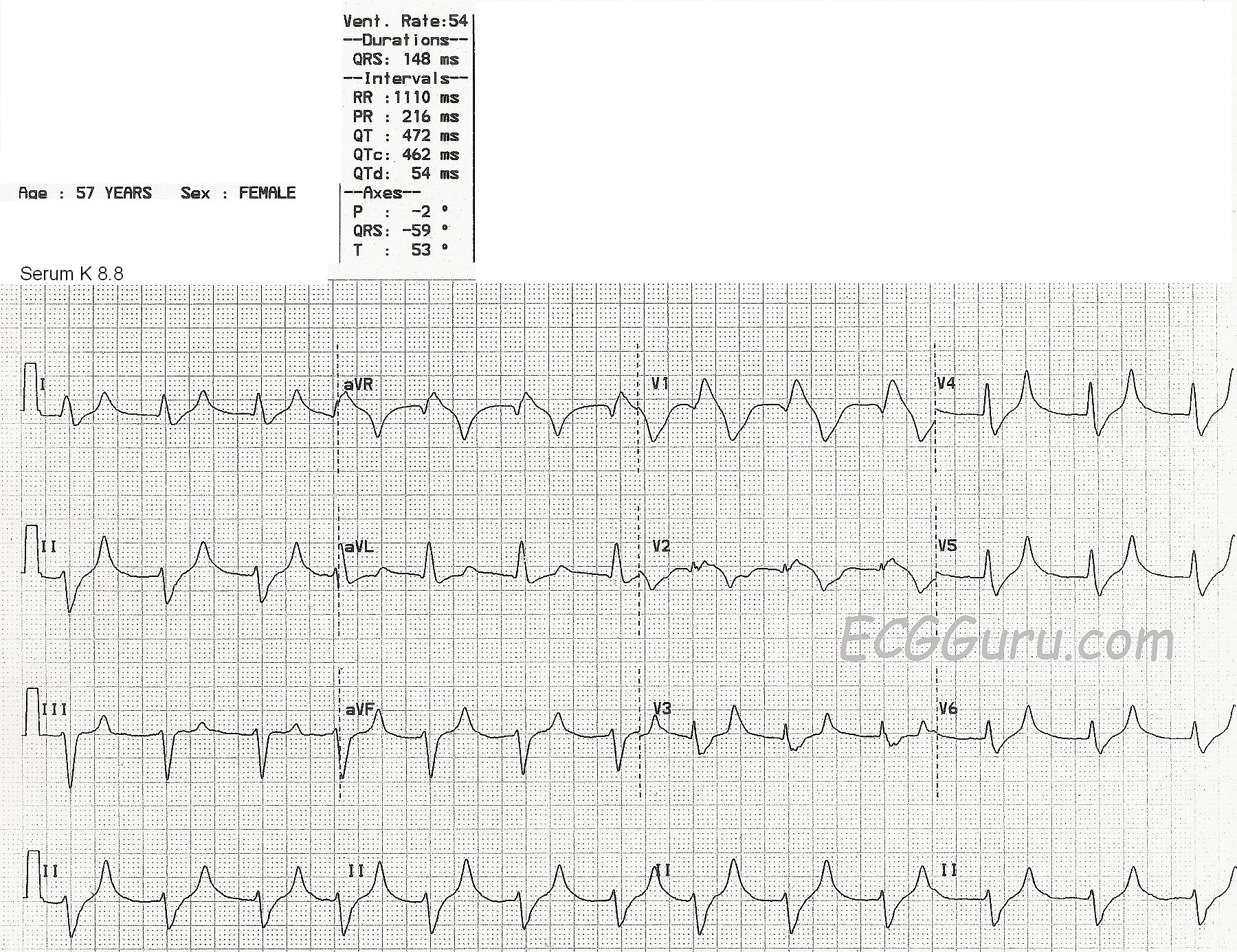
This ECG is from a 57-year-old woman with extreme weakness. We do not know her medical history or complete lab results, except that her serum potassium level was 8.8 mEq/liter at the time of this ECG.
This ECG shows a fairly regular rhythm at about 75 bpm, with a few early beats raising the rate slightly. (Even though the machine's interpretation lists the rate as 54 bpm. The QRS duration is listed at 148 ms (.148 seconds), but it appears wider. It is difficult to see the excact location of the J point because the QRS slurrs into the ST segment. Even though the ECG machine reports a P wave axis and a PR interval, P waves are not visible.
The QRS morphology appears to be an atypical right bundle branch block and left anterior hemiblock pattern. The T waves in leads I, II, aVF, and V3 through V6 are narrow, tall and peaked.
Potassium is primarily an intracellular electrolyte. It is necessary for proper electrical functioning of the heart. Extracellular serum potassium can rise due to renal failure, or taking potassium supplements, potassium-sparing diuretics, or ACE inhibitors. Occasionally, serum K levels may be artificially elevated by drawing the blood with too much syringe pressure, or using too small a needle, as the red blood cells can be damaged and release intracellular K into the serum.
ECG signs may vary among people with hyperkalemia, but in general:
Serum K levels of 5.5 mEq/L or greater can cause repolarization abnormalities like tall, peaked T waves.