
Submitted by Dawn on Mon, 07/02/2012 - 22:18
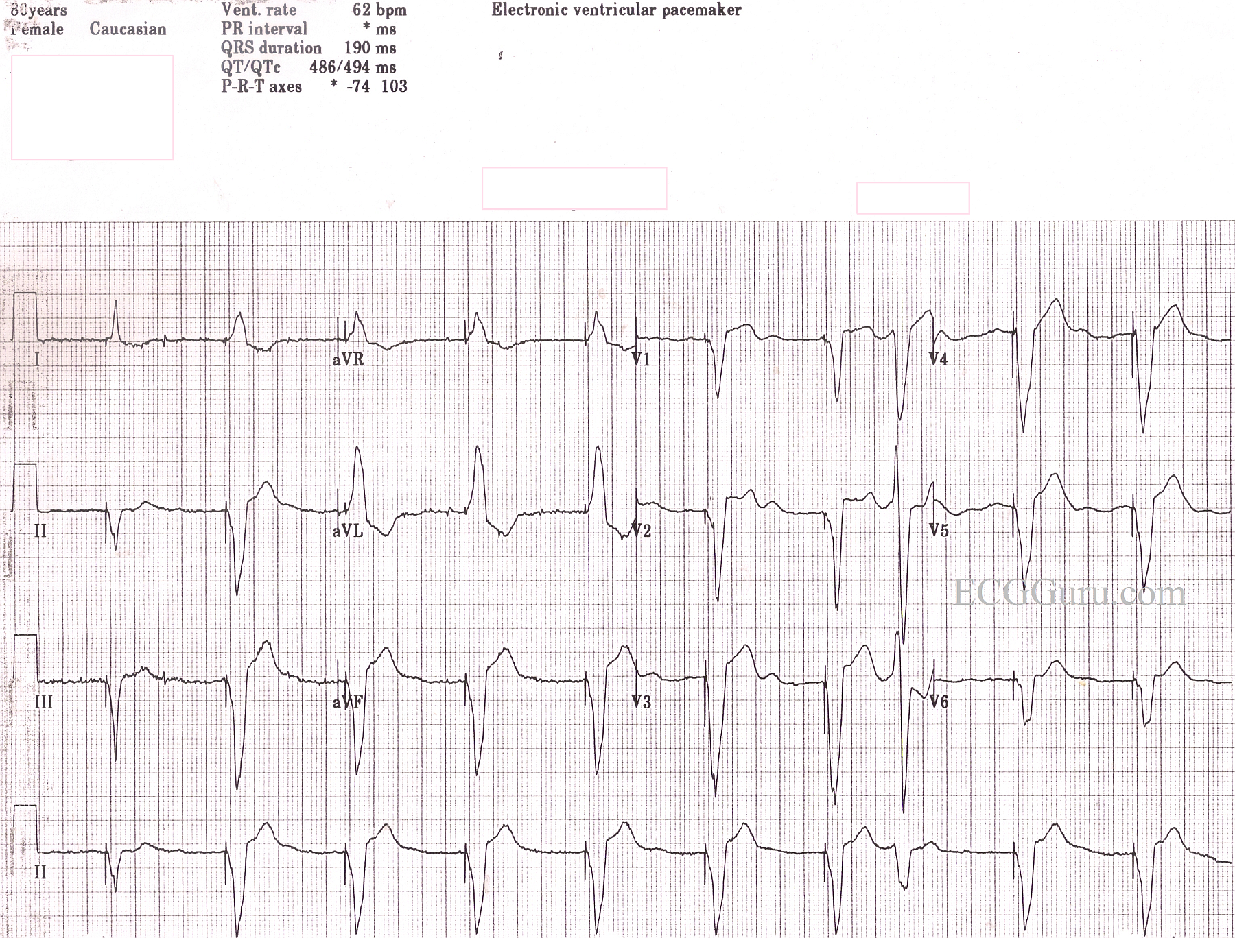
This is a good teaching ECG for beginners just learning to recognize paced rhythms. Spikes are not always this easy to see! All the characteristics of pacing are here, including spikes, of course. There are wide QRS complexes, indicating only one ventricle is being paced. The impulse spreads slowly from the right ventricle to the left. The axis is leftward because the left ventricle is being paced from the right ventricle, which is located down near the diaphragm. The rate is typical of a paced rhythm. This patient has an AV sequential pacer, but the underlying rhythm was atrial fibrillation with a controlled ventricular response. The A Fib is inhibiting the pacer from making P waves. For your more advanced students, the first beat is narrow, which may represent fusion, and the eighth beat is a native beat - probably a PVC. When interpreting a paced rhythm, it helps to have a long rhythm strip or serial 12-leads. But the best way to determine if the pacemaker is functioning as programmed is to have it interrogated by the manufacturer's representative. Remember, when the QRS is wide, discordant ST changes are normal - that is, negative QRS complexes will have ST elevation, and positive QRS complexes will have ST depression. To see a paced rhythm with ST elevation M.I., search "Paced Rhythm with M.I.".
Related Terms:
Rate this content:
All our content is FREE & COPYRIGHT FREE for non-commercial use
Please be courteous and leave any watermark or author attribution on content you reproduce.