Displaying 1 - 5 of 5
Dr A Röschl's picture

An Interesting Holter Strip

Here you can see a long rhythm strip from a Holter ECG, written at 25 mm/s. On the left, a sinus bradyarrhythmia can be seen first, followed by an atrial tachycardia. After a few beats this changes back into a sinus bradyarrhythmia. Then follows a short VT over 3 beats, after 1 sinus node beat then a ventricular couplet. Sinus bradyarrhythmia again at the end.

Dr A Röschl's picture

Ventricular Tachycardia In A Patient With CAD And An Old Inferior M.I.

Broad complex tachycardia in a 78-year-old patient with coronary heart disease (CHD) and an old inferior myocardial infarction. Why is this a ventricular tachycardia (VT) and not a supraventricular tachycardia (SVT) with aberrant conduction? Broad complex tachycardia is generally about 80% likely to be ventricular in origin. However, in a patient with CHD and a history of myocardial infarction, this likelihood increases to about 90%. What other clues are there? The tachycardia initiates with a premature QRS complex without a preceding/premature P-wave.

Dr A Röschl's picture

Ventricular Tachycardia After Inferior Myocardial Infarction

76-year-old man, with a history of inferior wall myocardial infarction. He experiences recurrent episodes of brief palpitations, often lasting only 5-15 seconds. In this ECG, at the beginning, the last part of a wide-complex tachycardia is visible. After 2 sinus beats, another wide-complex tachycardia begins (with the same QRS configuration).

Dawn's picture

Non-Sustained Ventricular Tachycardia

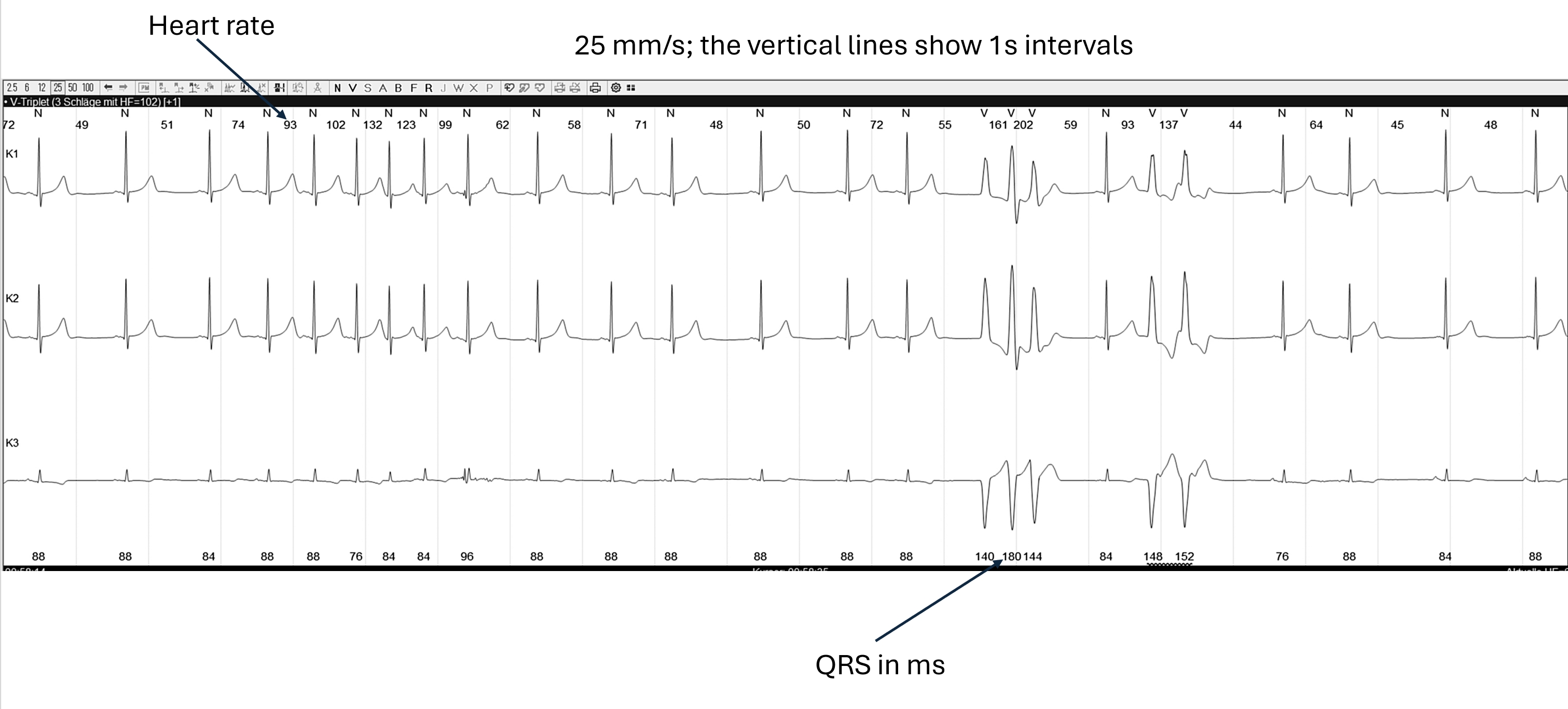
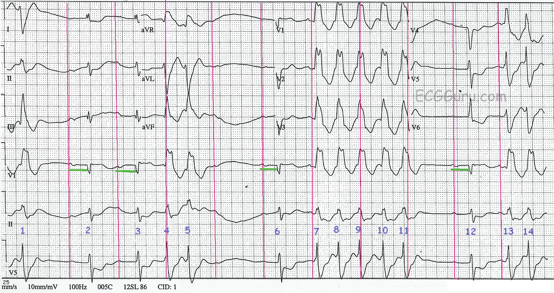
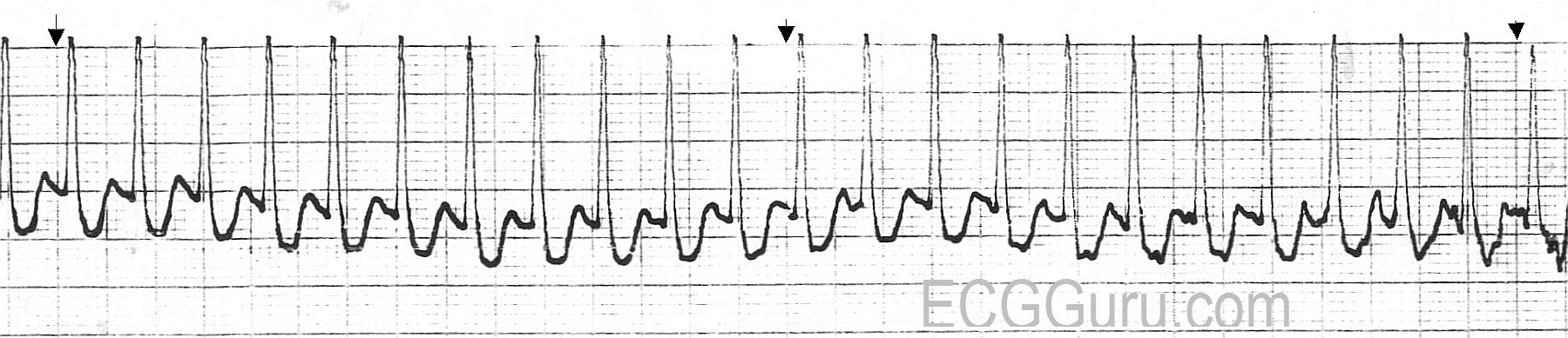
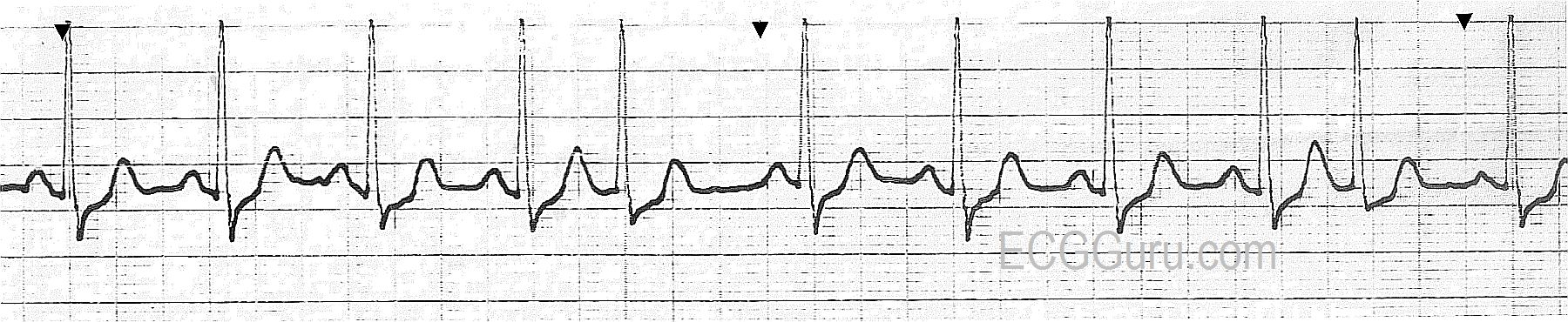
This ECG was obtained from a 45-year-old man who was experiencing palpitations and lightheadedness, which he originally attributed to anxiety. There are short, but frequent periods of ventricular tachycardia, which are self-limiting.  This is called "NON-SUSTAINED VENTRICULAR TACHYCARDIA".  The underlying rhythm is sinus, with a remarkably long PR interval, and at least one episode of failure of the P wave to conduct, making "second-degree AV block, Type II" a possibility. It is difficult to thoroughly evaluate the underlying rhythm because it is not seen very often in this ECG. The rate of the underlying P waves is about 67 bpm.  The PR interval is .40 seconds (400 ms).  The "normal" QRS complexes are slightly widened, at about .10 sec (100 ms), which is typical of Type II AVB.  The ventricular QRS complexes are wide at .16 sec. (160 ms)

To assist you in using this tracing for teaching, we have also supplied a "marked up" version.  The P waves, both visible and hidden, are marked with red lines. The PR intervals are shown in the Lead V1 rhythm strip in green.  And the QRS complexes are numbered.  QRS complexes numbered 2, 3, 6, and 12 are sinus. The P wave AFTER QRS #5 is non-conducted.

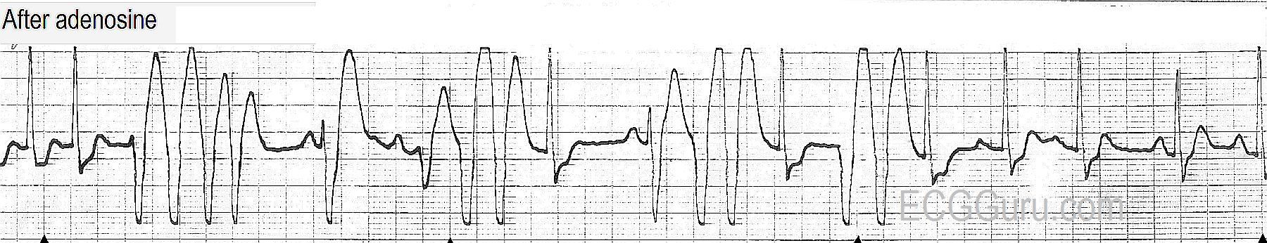
To review the differentiation of ventricular tachycardia from supraventricular tachycardia with aberrant conduction, go HERE.

Dawn's picture

ECG Basics: Paroxysmal Supraventricular Tachycardia Converted With Adenosine

Today, you get THREE strips for your basic classes!  The first shows a PSVT - paroxysmal supraventricular tachycardia at a rate of about 220/minute.  The QRS complexes are narrow, and the rhythm is regular.  In the second strip, we see the moment of conversion after a dose of 6 mg. of adenosine was administered by rapid I.V. infusion.  The re-entry cycle is broken, and the patient experiences many PVCs, including groups of two, three, and even four in a row.  Soon, however, the rhythm settles into sinus rhythm with PACs, and later, just sinus rhythm (not shown).  For your more advanced students, the second strip shows the PVCs interacting with the underlying sinus rhythm.  There are several fusion beats with varying degrees of fusion, and one can sometimes see a sinus P wave just before a PVC.  None of this is clinically significant in this patient, because the ventricular ectopy was a side effect of the treatment, and was self-limited.  It is a great strip for teaching, though! 

All our content is FREE & COPYRIGHT FREE for non-commercial use

Please be courteous and leave any watermark or author attribution on content you reproduce.