Displaying 1 - 10 of 19
Dawn's picture

Ask the Expert

Dr. Jerry W. Jones, MD, FACEP, FAAEM

QUESTION:

Dr. Jones, what advice do you have for teaching ECG beginners? 


Today’s expert is Dr. Jerry W. Jones, MD, FACEP, FAAEM
 

Jerry W. Jones, MD FACEP FAAEM is a diplomate of the American Board of Emergency Medicine who has practiced internal medicine and emergency medicine for 35 years. Dr. Jones has been on the teaching faculties of the University of Oklahoma and The University of Texas Medical Branch in Galveston. He is a published author who has also been featured in the New York Times and the Annals of Emergency Medicine for his work in the developing field of telemedicine. He is also a Fellow of the American College of Emergency Physicians and a Fellow of the American Academy of Emergency Medicine and, in addition, a member of the European Society of Emergency Medicine. 

Dr. Jones is the CEO of Medicus of Houston and the principal instructor for the Advanced ECG Interpretation Boot Camp and the Advanced Dysrhythmia Boot Camp. 

ANSWER: 

Even in my advanced classes I begin with "normal" ECGs. Throughout my residency in internal medicine, I was never up at 3 am wondering if an ECG was ABNORMAL ... I was always trying to decide if a finding was really NORMAL instead.

Here are a few of my thoughts...

A biphasic P wave in V1 is basically the norm. Even when there is only a monophasic deflection, it's usually because the other half of the biphasic deflection is isoelectric.

In my advanced courses we always begin with a normal tracing and I have all the participants measure the R-R intervals with ECG calipers to demonstrate that there is often considerable variation in the rhythm and that there is very rarely a perfectly regular sinus rhythm (and when there is - it's only for a few moments!). This comes in handy occasionally in deciding whether a tachycardia is sinus or not.

I often find that beginners have the impression that the R waves in the precordial leads increase in size from V1 through V6 - and that should never be the case in a "normal" ECG. Typically the tallest R wave peaks at V4 or V5. Because the V6 electrode is the furthest of all the regular precordial leads from the surface of the heart, it actually diminishes in amplitude. When the R wave in V6 is the tallest across the precordium, it means that the heart has enlarged enough to extend its surface a lot closer to the V6 electrode. That alone is a very good indication of cardiac enlargement.

One other thing I would really emphasize to a newbie is that the ST segment should rise gently into the upslope of the T wave and that there should never be a perceptible angle indicating where the ST ends and the T wave begins - it should be smooth and without a discernible margin. And the T wave should always be asymmetrical - NOT symmetrical. However, when the downslope of the T wave returns to the baseline it CAN create a noticeable angle. 

I hope some of these comments help you teach those who are just beginning to read ECGs.

Dawn's picture

Ask The Expert

Dr. Ken Grauer

QUESTION: 

Can you provide some guidelines on how to convey the large body of information associated with clinical evaluation and management of cardiac arrhythmias from a primary care perspective? 

Today’s Expert is the ECG Guru’s Contributing Expert, Dr. Ken Grauer. 

KEN GRAUER, MD is Professor Emeritus (Dept. Community Health/Family Medicine, College of Medicine, University of Florida in Gainesville).

Dr. Grauer has been a leading family physician educator for over 30 years. During that time he has published (as principal author) more than 10 books and numerous study aids on the topics of ECG interpretation, cardiac arrhythmias, and ACLS (including an ongoing Educational ECG Blog) . Dr. Grauer is the Contributing Expert for the ECG Guru. 

ANSWER: 

The topic of evaluating the patient with a cardiac arrhythmia – and then formulating an optimal approach to management is HUGE. It encompasses assessing both symptomatic and asymptomatic patients – determining if an arrhythmia is truly present, and if so, whether the arrhythmia is worrisome or benign – and then deciding on whether drugs, lifestyle changes, or referral for specialized EP (electrophysiologic) evaluation is in order. 

I have developed a 3-part (less than 90-minute) video series that addresses this tremendously important clinical topic from start to finish. Included in these videos are assessment of the patient, arrhythmia monitoring methods, when to refer, and targeted discussion of the most commonly encountered arrhythmias in primary care. These include ectopic beats (PACs; PJCs; PVCs) – ventricular arrhythmias (nonsustained vs sustained VT occurring in different clinical settings) – bradycardias (diagnosis of Sick Sinus Syndrome plus indications for pacing) – MAT – PSVT/AVNRT – Atrial Flutter – and Atrial Fibrillation. 

Links to these 3 Videos – plus LOTS of additional relevant information (including pdf excerpts available for free download from my ECG and ACLS ePubs on specifics of arrhythmia diagnosis) – is now all available for use on my new clinical arrhythmia webpage, www.fafpecg.com. I hope this material is helpful to you in your teaching! The beauty of these videos is that content is appropriate and understandable for primary care providers of virtually any level or degree of training – and that by assignment, learning can be entirely self-instructional OR under your direct guidance and instruction.  

P.S. I am still finishing a Power Point Show (without audio) that you can use if you choose to teach this subject yourself using my slides. This should be completed soon. The rest of this web page is finished and READY for use. Your comments and feedback is WELCOME!


If you have a question for one of our Experts, please email your question to:  [email protected]

Dawn's picture

Ask The Expert

Darlene Hutton, RN, BScN, MSN

Question:  As a fairly new educator in the telemetry/medical unit or Emergency Department, what steps would you suggest taking in helping new nurses to the unit understand ECG Interpretation?


Today's Expert is Darlene Hutton, RN, BScN, MSN

Darlene Hutton has worked in CCU, ICU, PACU, and Emergency as a bedside nurse, educator, and manager. She is currently working as a Clinical Research Manager at Rouge Valley Metabolic Research Associates in Toronto. Darlene also is an Educational Consultant providing workshops, seminars, and conferences on such topics as ECG Interpretation, Dysrhythmia Interpretation, Acute Coronary Syndromes and Cardiology Drugs. She is also an ACLS Course Director and runs courses throughout the province. Her company, QRS Educational Services, founded 18 years ago, provides education to nurses and other health care professionals throughout Canada. She is also the Department Head for Emergency Preparedness in the O.R. in the American College of Plastic Surgical Nursing journal.

 [email protected]

(905) 706-3301     www.qrs-education.com     www.facebook.com/qrs.educationservices


ANSWER:

As an independent educator for the past 20 years, I have had the incredible pleasure of spending time with novice to expert nurses in their respective field from a variety of different hospital settings.  My response to this question comes from what I have learned from each of these individuals over the past 2 decades.

We know that the “expert” practitioner is sometimes not the best educator because of their own challenges in bringing the information to a basic level of understanding.  What makes an educator invaluable is their ability to bring the most advanced concepts to the level of understanding for the individual and tailor the education to that level.

A novice nurse entering into one of these units will likely have had some courses in rhythm interpretation as a basic entry level requirement.  However, I find that many of these nurses have not had adequate preparation time in being able to apply a systematic approach to assessing the 12 Lead ECG. When working in these types of units, promptly assessing the ECG is essential to ensure there is no time delay in notifying the physician of anything urgent.

I apply a step-wise approach in ensuring that the basics are understood before moving on to the more intricate aspects of the ECG.

The basics that I assess are:

1. Ensure the nurse understands what the P, QRS, and ST represent and their significance, if abnormal. Most importantly, I ask them to point out examples of ST elevation and depression on various ECGs.  You would be surprised at how many people do not know this very core concept and before discussing treatment management and complications, this basic concept needs to be well understood. If you only have 30 minutes to spend with a nurse on this particular day, this would be the priority, in my opinion.

2. The next step would then be to assess their knowledge and discuss what’s happening with ST elevation/depression, including the treatment strategies. This can be achieved in 30 minutes to an hour.

3. Third: anticipated complications. I can’t tell you how often I hear nurses want to give a saline bolus to a hypotensive patient experiencing an anterior wall MI.  They do not understand the pathophysiology and their actions will subsequently worsen the situation.  Apart from cardiac arrest occurring with any MI, I expect that everyone understands the ‘expected’ complications associated with the inferior and anterior wall MI. For this, I keep it very simple so retention of information remains. “The heart is comprised of 2 pumps. If the right pump fails and backs up, what will you notice? Will the chest be wet or dry? (about 1/3 of the group will say wet, which is incorrect). If the left pump fails and backs up (such as in an anterior MI), where does it back up to? Will the chest be wet or dry?” By keeping it simple, the concepts are easier to understand and retain.

4. In many of the hospitals, there is an annual recertification of the ECG basics and this is another opportunity as an educator to assist those who may be struggling. 

5. I also provide staff with reference tools so they can help themselves learn at their own pace. On my educational website: www.qrs-education.com , there are reference links and tips under the QRS Tips tab as well as our manual which you can read about in the Products page. This manual has taken the complex concepts to a more manageable level. I also promote Dawn’s website (www.ecgguru.com ) as another invaluable tool. Two examples of interactive links are:  www.practicalclinicalskills.com  and www.skillsstat.com 

I hope this has helped. Thank you.

Darlene Hutton

President, QRS Educational Services

www.qrs-education.com  


 IF YOU HAVE A TEACHING QUESTION FOR OUR EXPERTS, PLEASE EMAIL TO [email protected]


Dawn's picture

Teaching Tip: A Series of ECGs Can Tell A Story

When teaching ECG, I always try to make the ECG interpretation have some practical context for the student.  Why study squiggly lines, if they don't mean something to our care of our patients?  Even putting a simple scenario (actual or invented) with an ECG can make it more relevant for your students.  A series of ECGs taken as the patient undergoes changes, is especially helpful.

Dawn's picture

Teaching Tip: 12 Leads are Better Than One (Or Three)

Years ago, I was tasked with introducing 12-lead ECG interpretation to firefighter/paramedics who had been using ECG for rhythm monitoring for years.  Some were eager to add to their skills, others - not so much.  The feeling was, we have been doing just fine as we are.  When finally convinced that they could interpret STEMI with a 12-lead, many were content to use the 12-lead ECG only for that.  

To illustrate to students the great value of multi-lead assessment, I devised a little "quiz".  I showed the students ten to twelve short rhythm strips, like you see here.  All were cropped from 12-lead ECGs.  I asked my class to interpret the strips as they would if they were taking an ACLS class.  Usually, all did fine, or so they thought.  When shown the 12-lead ECGs the strips were taken from, EVERY student changed his or her mind on EVERY ECG.  The lesson is:  sometimes what we are looking for shows up in some leads and not others.  You can find this illustrated hundreds of times just in the ECG archives on this site.  I will supply some ECGs here on this page over the next few weeks that you could use to show your own students the value of "multi-lead assessment".  

What started as a hard-sell turned out to be a fun exercise.

The ECG shown here is of a patient in V Tach.  There are several strong signs that this is V Tach, including the wide QRS complexes, lack of associated P waves, "backward" axis, also called extreme right axis deviation (Leads II, III, and aVF are all negative and aVR is positive), and V6 is negative.  For more review of the differential diagnosis of wide-complex tachycardias, go to our Ask the Expert answer from Jason Roediger.  This LINK willl take you to Dr. Grauer's informative webpage where he offers a step-by-step guide to differentiating the WCTs.

 The focus of THIS lesson is that, while the patient is in V Tach, and it is in every lead, the tell-tale signs are harder to see in some leads than others.  Remember to show your  Remember to share with your students that the channels of the ECG (in this case three) are run simultaneously, so that the same heartbeat is seen several times - once for each channel.

Dawn's picture

Ask The Expert

Question:  I teach ECG, but I don't work in a cath lab. What important points would you, as an Interventional Cardiologist, like for me to convey to my students?
Today'sExpert is Dr. Stasinos Theodorou.   Dr. Theodorou
 

Dr. Theodorou completed his medical training in 2002 at the University of Leicester medical school, United Kingdom.  Following his yearlong pre-registration practice he completed his three year general / internal medicine training which resulted in achieving membership of the Royal College of Physicians of the United Kingdom (MRCP UK).  He further specialized as a cardiologist within the West Midlands, completing a period of five years specialist training, in the University Hospital of Birmingham, and University Hospital of Coventry and Warwickshire with a subspecialty in interventional cardiology.  After completing the necessary official examinations in Ioanina, Greece, he was accredited with the specialty title in Cardiology and returned to Cyprus in 2011.

Currently, Dr. Stasinos Theodorou is a member of the Cyprus Medical Association, as well as the Cyprus Society of Cardiology.  He is also a member of the European Association of Percutaneous Cardiovascular Intervention (EAPCI), and the British Cardiovascular Intervention Society (BCIS).  During his training he has been academically active and has published several scientific articles in well-respected medical journals. He has also participated in medical research trials through his different posts in UK University Hospitals.

 For more from Dr. Theodorou, check his website, Limmasol Cardiology Practice at http://www.cardiolimassol.com and his very interesting FaceBook page, http://www.facebook.com/#!/LimassolCardiologyPractice. 

ANSWER:

The job of the emergency cath lab team is very important, stressful and tiring, but above all rewarding. It’s really a great feeling finishing a case with your patient well and smiling, and his/her family being relieved and thankful. The cardiologist and the cath lab team, however, are the last link in a chain of health professionals to actually be involved. In order for the entire system to function productively every single one of these links has to be in place, doing their job precisely and timely.  

The patient first of all has to seek attention, let’s say present to casualty. For this to happen there should be some sort of awareness campaign within a community to educate public and warn about the typical symptoms etc. If a patient does not present early it might be catastrophic. 

The next link in the chain is the paramedic and / or the casualty staff (if the patient self presents instead of calling 911) This is probably the most crucial step in the patient’s journey into the lab and a lot of the credit for saving a life should be attributed to these amazing people who are constantly on the watch!  

As an interventional cardiologist I want to be called out at any time, ON TIME with as little as possible time wasted. From the moment I receive that call I have 90 minutes (preferably 60 according to recent guidelines) to get that artery unblocked and in these cases “time is muscle”. For this exact reason I expect paramedics, nurses, casualty officers or internal medicine physicians to recognize the trigger points on an ECG and MATCH it to the individual patient’s clinical scenario and if appropriate CALL US IMMEDIATELY! Of course there will be false alarms but I would prefer to have those rather than a patient with a MI sitting on a trolley in ED while his/her myocardium dies. 


So, when should the cath lab team be alerted? 

Starting from the basics one should begin with the patient’s presentation and nature of complaint ( After all the patient is not just an ECG!)

Is the presenting complaint typical of an MI? Is the nature of the pain cardiac, or could it be something else?

Getting it wrong at this stage may result in missing other important diagnoses (Pulmonary embolism, aortic dissection, pneumothorax etc) and alerting the cath lab will simply waste crucial time until the appropriate treatment is delivered. If the symptoms are atypical reconsider. 

An ECG must be obtained within 10 minutes of the first medical contact.


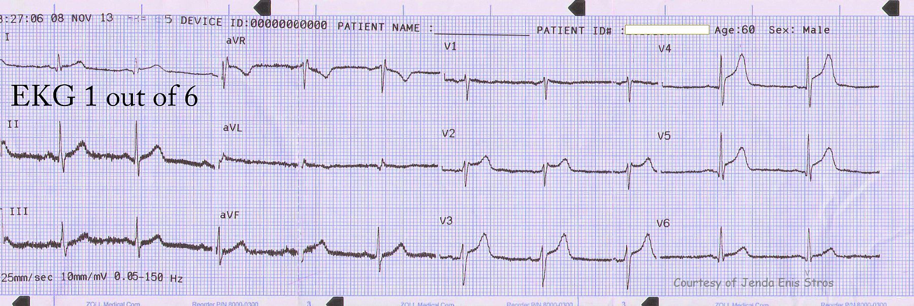
If the symptoms are typically cardiac the following ECG patterns should instantly trigger the team: 

1.      ST segment elevation measured at the J point in two contiguous leads. Certain specific voltage criteria apply depending on issuing body (AHA/ACC or ESC) but generally speaking anything more than 0.1mV should be considered as suspicious.

2.      ST segment depression in leads V1-V3 (> 0.05mV) with positive terminal T-waves or associated with dominant R-waves in V1-V2. This pattern is suggestive of a posterior STEMI and this can be confirmed by obtaining posterior leads (V7-V9).

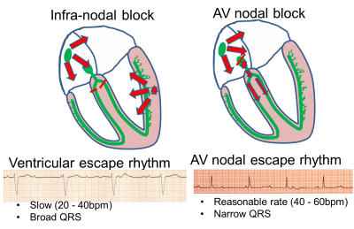
3.      New onset LBBB (If no previous record consider as new, unless proven otherwise)

4.      Isolated ST segment elevation in aVR (usually signifies left main coronary occlusion when associated with widespread ST depression in other leads)


ECG patterns that can prevent diagnosis and should be considered highly suspicious if accompanied by typical symptoms of ongoing ischaemia: 

1.      RBBB shouldn’t hinder the diagnosis of STEMI but patients with MI presenting with RBBB have a poor prognosis.

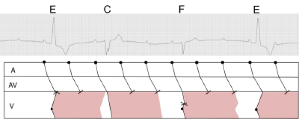
2.      Ventricular paced rhythm. If possible and not time consuming reprogramming the device to evaluate the intrinsic pattern should be considered.

3.      Pre-existing LBBB can also impede the diagnosis of an acute MI. Often with acute MI there are marked ST/T changes on top of the underlying LBBB. 

Conditions that mimic acute ischaemia on a 12 lead ECG.

These conditions require careful assessment before excluding acute myocardial ischaemia and a cardiology consultation should be requested. 

1.      Left ventricular aneurysm as a result of an old, full thickness MI. This is associated with Q-waves in the affected territory.

2.      Left ventricular hypertrophy. This could be due to primary hypertrophic cardiomyopathy, severe aortic stenosis or hypertension.

3.      Pericarditis  or myopericarditis) usually produces a typical appearance of ST elevation (often called saddle shaped”) which does not fit to a coronary distribution pattern.

4.      Brugada syndrome. This is a genetic disease characterised by an abnormal ECG and predisposes to sudden cardiac death.

5.      “Takotsubo” or stress-induced cardiomyopathy causes ST elevation with a specific LV pattern (apical ballooning) with normal coronary arteries.  Takotsubo, Typical appearance of LV during systole of LV

6.      Hyperkalaemia can sometimes cause ST elevation with tall T-waves

7.      Sub-arachnoid haemorrhage can cause ECG abnormalities including ST elevation.

8.      Pulmonary embolism.

9.      Repolarisation abnormalities.

10.  ST segment elevation has also been reported in various intra-abdominal conditions, including acute pancreatitis and large hiatus hernia causing cardiac compression.


In the presence of typical symptoms, without any of the above patterns the alarm should not be triggered but that does not mean reassurance.  If the initial ECG is normal look for hyper-acute T waves which precede ST elevation.


If there is ischaemia in the form of ST/T changes the patient should be treated accordingly (Antiplateletes, nitrates, low molecular weight heparin +/- Glycoprotein 2b/3a inhibitor) and a 12 lead ECG should be repeated at regular time intervals. If at any given point the ECG shows one of the above trigger patterns, the cath lab team should be called. In these high risk, intermediate cases, continuous 12 lead monitoring is preferable but not always available.


With intractable typical symptoms of ongoing ischaemia the possibility of occluded non-dominant circumflex artery should be considered which sometimes does not result in substantial ECG changes. In this case I personally feel that the decision to take the patient to the lab should be made by a cardiologist. 


If the symptoms are very atypical (sharp pain, worse on inspiration, radiation between the scapulae) other diagnoses should be excluded even with a suspicious ECG. A chest X-ray takes less than 10 minutes and can give you a lot of information. At the end of the day I don’t want a patient with tension pneuothorax or dissecting thoracic aneurysm on the cath lab table! (And I did have!!) If the suspicion for aortic disease is high (clinically with the classic signs) a CT scan should be done first. 


If there is any diagnostic uncertainty a quick, bedside echocardiogram will often provide the decisive information to guide your next move. It may demonstrate regional wall akinesia consistent with MI or point to a different diagnosis such as aortic dissection or even pericarditis. 


When you have covered all the above and you still feel hopeless, not knowing what on earth is wrong with your patient who is in pain with a normal ECG check the following:

1.      Amylase. You would be surprised how many times I diagnosed pancreatitis as a cardiologist!!

2.      D-dimers. A pulmonary embolism is subtle but deadly and not always easy to diagnose.

3.      Surgical opinion if there is any suspicion of gall stones or other upper abdominal pathology (Gastritis, oesophagitis etc)

4.      Creatinine Kinase, CKMB, Troponin. It’s never late to diagnose an MI! Your local cardiologist wouldn’t hold you responsible in the absence of ECG changes.

5.      Never lose hope!! Do all of the above again if necessary.


   RCA with 100% occlusion       RCA post intervention               Culprit clot


In fact the easiest case scenario for paramedics and casualty (emergency) personnel is a patient with a straightforward, typical, barn door MI for you to simply pick up the emergency phone and call us! We will do the rest starting from getting a diagnostic angiogram, deciding which is the culprit lesion, aspirating the clot, dilating the stenosis and implanting a stent! And yet the most important job of all is that CALL, without which none of the above would ever happen!


Dawn's picture

Ask The Expert

QUESTION:  Do you have any "tricks of the trade" for teaching complex ECG topics?

Our expert today is Dave Richley.            

Dave Richley worked as a clinical cardiac physiologist, in echo, pacing, Holter etc, for over 30 years in various hospitals in North-east England, but for the last few years he has been employed almost entirely in an educational capacity. He teaches undergraduate and postgraduate clinical physiology degree programmes at Sunderland and Newcastle Universities and is currently engaged in a project with the British Heart Foundation, the UK’s leading heart disease charity, to deliver courses in ECG interpretation to cardiac nurses. His particular interest within the field of electrocardiography is complex bradyarrhythmias and conduction disturbances.

"I believe that in this era of high-tech multi-modality imaging, the subtle and complex art of ECG interpretation has become something of a forgotten art and my mission is to see electrocardiography restored to its former and rightful pre-eminent position in diagnostic cardiology!" – Dave Richley


ANSWER:


For teaching all but the simplest of topics I am increasingly using pictures. Verbal explanations of ECG phenomena, to those who are not that familiar with the sequence of electrical events and the anatomy of the conduction system in the heart, can be difficult to understand. I have therefore produced a simple picture of the heart on which I superimpose various arrows and other shapes, as appropriate, to help me illustrate various phenomena. Below is an attempt to explain with the help of simple diagrams the different ECG manifestations of complete AV block at the level of the AV node and below the AV node in the ventricles. I have no artistic ability and no special software – I make all my pictures within Powerpoint using the standard drawing tools available.


To explain more complex arrhythmias, I need more complex pictures, and I find that laddergrams serve this purpose very well. Convoluted verbal explanations of complicated arrhythmias may just cause bafflement. The ECG itself is merely a depiction of the sequence of depolarisation of the muscle mass of the atria and ventricles; crucially it does not explain  conduction – or lack of conduction – directly; these phenomena must be inferred or deduced, and the laddergram is excellent for illustrating the origin and direction of propagation (or failure of propagation) of electrical impulses through the heart. Those inexperienced in ECG tend to assume that if a P wave is seen immediately before a QRS, this indicates that the atrial impulse is conducted to the ventricles. Of course, this is usually the case, but in AV dissociation this relationship between P wave and QRS may be purely coincidental. This is a phenomenon that can be illustrated very clearly with a laddergram.  I have tried to use the laddergram below to illustrate what is happening in a case of high grade AV block, with the blocks of pink representing ventricular refractoriness. There are no rules governing laddergrams, just conventions, and I think that people should be free to construct them as they wish in order to illustrate the explanations they are trying to convey. The only absolute requirement is that the deflections of the ECG must be exactly simultaneous with the corresponding lines on the laddergram. There are several ways of constructing laddergrams; my own method is to produce them within Powerpoint by inserting shapes – mostly just straight lines – and copying and pasting them into the correct positions. With practice, this can be done quite quickly.


Dawn's picture

Ask The Expert

 QUESTION: How do you explain the difference between "AV block" and "AV dissociation" to your students?

Our Expert today is Christopher Watford, BSc, NREMT-P 

Christopher began in EMS as an EMT on a volunteer industrial fire brigade at GE's Global Nuclear Fuels facility in Wilmington, North Carolina. He has worked there as a Lead Software Engineer since 2001 and currently is a Captain on the fire brigade. Outside of his day job, he volunteers as a Paramedic and Field Training Officer for Leland Volunteer Fire/Rescue where he also serves on the board of directors.Through Cape Fear and Brunswick Community Colleges heteaches continuing educat ation for all levels of providers. He also is an associate editor for the EMS 12-Lead Blog and Podcast, presenting electrocardiography case studies for pre-hospital personnel. 

Christopher's excellent blog can be found at My Variables Have Only Six Letters.  His contributions to EMS 12-Lead can be found at this link.


Answer:

I think the first step in understanding the difference between an  atrioventricular block and atrioventricular dissociation is to have a  firm understanding of physiological and pathological conduction.  The most common example of this is a non-conducted premature atrial  contraction (PAC). If an atrial stimulus arrives early enough at the  atrioventricular node (AVN), while it is still refractory, forward  conduction will be blocked. Likewise in atrial flutter, you typically  see one ventricular activation for every two F-waves, due to the  physiological rate limiting by the AVN. However, as this is due to the physiological function of the AVN we would not consider this a block!

 In higher degree AV blocks, we encounter a pathological decrease in  conduction and so we label non-conducted stimuli as "blocked". Type I  and Type II AV blocks provide visual confirmation of pathological conduction as you have examples of both conducted and non-conducted stimuli.  However, in the case of a presumed complete AV block, it is important  that you look at whether the atrial impulses were blocked or simply not conducted. With monomorphic ventricular tachycardia you may see  uncoordinated atrial and ventricular impulses on the ECG. In this case  the ventricular rhythm and the atrial rhythm "compete" for access to  the AV nodal tissue. There is no "AV block" present, instead we say they are "dissociated" from the ventricular rhythm. More specifically,  we say that the atrial rhythm is dissociated from the ventricular rhythm due to usurpation. Best illustrating the competitive nature of two rhythms during dissociation are capture or fusion beats.

 Therefore when classifying dyssynchrony between the atria and ventricles, students should look to see whether conduction blocked due to pathological processes or because the AV node is appropriately refractory.


Dawn's picture

Ask The Expert

QUESTION:  Do you have some "tricks of the trade" to share with other ECG instructors?


Our expert today is Darlene Hutton, RN, BScN, MSN

She has worked in CCU, ICU, PACU, and Emergency as a bedside nurse, educator, and manager. She is currently working as a Clinical Research Manager at Rouge Valley Metabolic Research Associates in Toronto. Darlene also is an Educational Consultant providing workshops, seminars, and conferences on such topics as ECG Interpretation, Dysrhythmia Interpretation, Acute Coronary Syndromes and Cardiology Drugs. She is also an ACLS Course Director and runs courses throughout the province. Her company, QRS Educational Services, founded 18 years ago, provides education to nurses and other health care professionals throughout Canada. She is also the Department Head for Emergency Preparedness in the O.R. in the American College of Plastic Surgical Nursing journal. 

[email protected] 

(905) 706-3301 

www.qrs-education.com 

www.facebook.com/qrs.educationservices


Answer:

I sure do have lots of tricks to help me retain what I learn and I pass these tricks to those that I spend time teaching. The reason I have developed these tricks is because I have found that I best learn from visual demonstration and KISS principles, not only that; but, if we make things too complicated from the get-go, people won't want to learn the concepts.


Trick #1: Using the RIRI approach to ECG assessment. The acronym RIRI reminds us to check Rate, Intervals, Rhythm, and Ischemia/Infarction. We need a fast, reliable method to systematically look at and ECG. I can't tell you how many times people rush to checking out the ST segment elevation which is plainly there and overlook the fact that the patient is in a bundle branch block or in a 3rd degree AV block. I have established this method for assessment over 15 years ago, teach is regularly, and use it myself. If you want to add a diminsion to the assessment, you can always throw in axis determination right when doing the Intervals; specifically the QRS interval.


Trick #2: Bundle branch blocks: Goodness, how this has gotten complicated. It really isn't. If there's a P wave in front of a wide QRS complex, that's a bundle branch block. Otherwise, if there is no P wave with a wide QRS complex, it would be a ventricular rhythm. Here comes the fun part, when you've established there is a bundle branch block, take a look at V1 on the 12 Lead ECG. If V1 is "upright", you have a "right" bundle branch block. If V1 is "down low", you have a "left" bundle branch block. There are many other quick, neat ways that also work, but it is difficult to explain without physically showing you. The most important part of determining a bundle branch block is knowing that a "left" will often mask an acute coronary syndrome; specifically an MI. Right bundle branch blocks don't tend to mask MIs nearly as much. So, forget the misconception about "bunny rabbit ears", or the confusion of "slurred S waves in the lateral leads". Keep it simple and you'll remember forever. Upright = right, Down Low = Left.


Trick #3: Axis is another area that has been so complicated and unnecessarily so. The first thing I share with my students is what axis is and the various non-critical causes of an abnormal axis: pregnancy, obesity, hypertrophy, pulmonary stenosis, neonates, pulmonary hypertension, previous MIs, and others. These are all conditions that do not occur overnight and do not cause concern for an abnormal axis. The second part that I share when teaching axis, is the "how". You need a simple, uncomplicated method to teach and remember axis. I was first taught, and still remember the 7 steps along with plotting, to determine the exact degree of axis. It dawned on me quite a few years after, that the cardiologists I worked with simply reported "axis normal, right, left, extreme". Why were we, perhaps still are, taught a method that few people retain? So, here's the simple, way I teach it for better retention: 


Normal:


Extreme:


Left:


Right:


Lead I


Lead II


When looking at axis, look at Leads I and II


Normal="2 thumbs up", both I and II's QRS are upright


Extreme="2 thumbs down", both I and IIs' QRS are negative


Left = "knees apart", that's a lewd way to sit


Right= "knees together", that's the "right" was to sit.


For those who want to understand axis, they need to understand when an abnormal axis is a concern. In my opinion, the cardiac patient who has just had an anterior MI with a right bundle branch block is the patient you need to be supervigilant on looking at their axis. Why? Well, if their axis suddenly changes in this situation, it's likely because that patient has just blocked off the last remaining electrical circuit in their ventricles and may very likely develop syncope and significant bradyarrhythmias.

If you're going to teach axis, you need to understand it well yourself and you need to understand hemi-blocks and bifascicular blocks, and teach these concepts in a non-complicated manner.


Hope these tricks help.


Dawn's picture

Ask The Expert

QUESTION:  How much time should the entry-level paramedic training program allot to ECG training, and how much should be rate and rhythm instruction vs 12-lead?

Our Expert Today is Marjorie Bowers, EdD, RN, Paramedic

Dr. Bowers has been involved in emergency medicine since 1968. Her wide range of experience includes emergency nursing, flight nursing, street paramedicine, and EMS education. Currently, she serves as a team member on both a federal Disaster Medical Assistance Team (DMAT) and a State Medical Response Team (SMRT).  Dr. Bowers holds a doctoral degree in higher education from Florida State University and a Master’s Degree in Educational Leadership from Florida Atlantic University. She is a Florida certified paramedic and Registered Nurse.  During her 26-year tenure at Indian River State College in Ft. Pierce, Florida, she received the State of Florida EMS Educator of the Year award, was an appointee to the Florida EMS Advisory Council, was chosen as one of only a few educators nationwide to serve on the Educational Standards Curriculum Revision Committee for the National Assoc. of EMS Educators and NHTSA. During this time, she also authored numerous self-studies for both EMT and paramedic programs and participated in successful state and national accreditation site visits. She currently is a team leader for paramedic program site visits for the Committee on Accreditation of Educational Programs for the EMS Professions.

Throughout her career, Dr. Bowers has assisted thousands of EMT and paramedic students to successfully  complete National and State of Florida certifying exams. She has developed and delivered presentations at numerous local, regional, state, and national meetings and conferences.


Answer:


That is a very good question and a very hard one to answer. The problem in answering this question is that paramedic programs vary significantly in length all across the US.  Also, some students will pick up ECGs very quickly where others struggle and may never be really proficient in them.

You can  probably teach basic rhythm interpretation in about 8-12 hours. That includes review of cardiac A&P. However, really learning ECGs takes practice. All instructors who have contact with paramedic students should quiz them from the time they learn rhythms until they finish the program.  

At the beginning of the 2nd semester, we reviewed ECG interpretation from 1st semester and then completed all of the rest of the rhythms. Again, once or twice a week, they would have ECGs on quizzes and always on tests.

In addition, students had ECG assignments to complete in clinical and ride time each semester. They could use textbooks or ECGs of real patients. By the time they finished the program, each student had IDs probably close to 1000 ECG strips (in class, lab, clinical, ride time)

12 lead probably could be taught in about 6-8 hours if you are including recognition and treatment of MI and angina and mimics. That would also include initial training on lead placement or review of lead placement if the students had already been trained on that skill.

During this class, I think it is important to keep practicing strips. Making each student “walk” you through the process of interpretation is helpful also.

Then again, practice, practice, practice. Keep going over the 12 lead strips in class, lab, clinical, and ride time.

Obviously, these times are relative to how long your program is.


For information about Dr. Bowers’s upcoming classes, contact Dr. Marjorie Bowers, Consulting, LLC at [email protected].

Pages

All our content is FREE & COPYRIGHT FREE for non-commercial use

Please be courteous and leave any watermark or author attribution on content you reproduce.