Displaying 1 - 4 of 4
Dawn's picture

Acute Anterior M.I. and Ventricular Fibrillation

The Patient:   This series of ECGs is from a 65-year-old woman who was complaining of a sudden onset of chest pain, nausea, and weakness. She stated that the pain increased on inspiration.  She reported a history of non-insulin-dependent diabetes mellitus (NIDDM). 

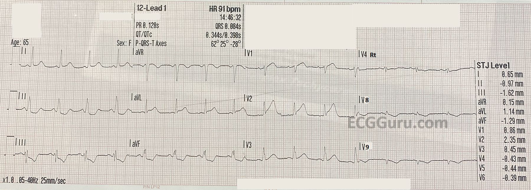
ECG No. 1, 14:46:  This ECG includes V4Right, V8 and V9 in place of V4, V5, and V6.  The rhythm is sinus at 91 beats per minute.  The PR interval is within normal limits, as is the QRS duration.  The QTc is WNL as well.  The frontal plane axis is also WNL.  The three standard chest leads show an early transition of R waves in V2.   There are noticeable ST and T wave abnormalities:

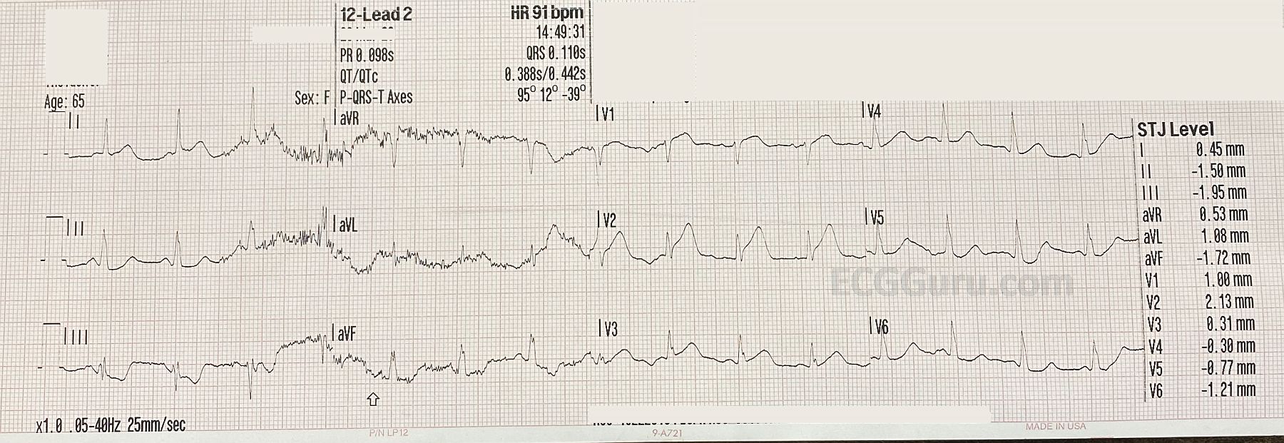
slight ST elevation in I and aVL with ST depression in II, III, and aVF.  In chest pain, possible M.I., STD should be presumed to be reciprocal in nature.  V1 has slight STE with a coved upward (frowning) appearance.  V2 has more noticeable STE, with a tall, wide-based T wave. This is called a “hyperacute T wave”.  We will have to evaluate V4 – V6 on ECG No. 2. 

V4 Right has no ST elevation, and V8 and V9 have ST depression (reciprocal to the anterior leads).  So far, we have all the signs of acute anterior wall M.I. 

Dawn's picture

Atrial Fib To Cardiac Arrest

A paramedic crew responded to the office of a local physician. A 61-year-old male presented with a one-week history of chest pain and shortness of breath. He had a previously undiagnosed atrial fibrillation with rapid ventricular response and left bundle branch block, but was alert. Shortly after transport commenced, the patient became unresponsive with Torsades de Pointes, which rapidly degenerated into ventricular fibrillation. The paramedic placed pads and defibrillated within one minute.  After two minutes of compressions, the patient had a fairly regular rhythm with return of spontaneous circulation.  Transport time was short.  On catheterization, the patient was found to have severe coronary artery disease, requiring coronary artery bypass graft surgery (CABG) A balloon pump was inserted in an attempt to strengthen him for surgery.

What is the rhythm?   The 12-lead ECG presented here shows atrial fibrillation at a rate of 138 per minute.  The rhythm is irregularly-irregular with no P waves.  Since the patient had not yet been diagnosed with atrial fib, obviously no therapy had been initiated to control the rate. There is a PVC near the end of the strip.

Dawn's picture

ECG Basics: Ventricular Fibrillation Converted With Defibrillation

A good example of ventricular fibrillation converted by electric defibrillation to what appears to be a sinus rhythm.  There is significant artifact in the post-conversion strip, but the last beat on the strip appears to have a P-QRS-T sequence.

Of course, V Fib presents without pulses, and must be defibrillated as soon as possible.  Best results are achieved by defibrillating a perfused heart, so if there is any delay from onset of V Fib, CPR should be performed to perfuse the heart prior to defibrillation.

Dawn's picture

ECG Basics: NIPS Procedure, ICD Test

This is a rhythm strip from a NIPS procedure (non-invasive programmed stimulaltion), which is a programming test for an implantable cardioverter defibrillator (ICD).  The test is done under light anesthesia, similar to that used for colonoscopy.   In this example, the patient is in normal sinus rhythm at the beginning of the procedure.  The pacemaker technician overdrives the patient's rate to observe the pacing function, then a stimulus is delivered to cause ventricular fibrillation (V Fib).  Initially, the ventricular rhythm is somewhat organized and coarse (V flutter), but it will rapidly deteriorate if not corrected.  Before it deteriorates, the ICD delivers a shock, and the patient's rhythm is restored.  In this example, bi-ventricular pacing was conducted for a few minutes before the patient resumed NSR.  The patient is then recovered from the anesthesia and discharged home.

For your students, this is a good example of the relative safety of shocking the well-perfused heart.  Although it is possible to put the heart into an intractable V Fib with this test, the ICD usually is able to convert the potentially lethal rhythm easily.  It is a good reminder that we need to perfuse the heart well before performing defibrillation on a person with unwitnessed cardiac arrest.

All our content is FREE & COPYRIGHT FREE for non-commercial use

Please be courteous and leave any watermark or author attribution on content you reproduce.