Displaying 1 - 7 of 7
Dawn's picture

Teaching Series - Tachycardia and Left Anterior Fascicular Block

This series of three ECGs is from a 60-year-old man who was brought to the Emergency Department after being involved in a motor vehicle accident.  No injuries were found, but the patient was severely intoxicated by alcohol consumption.  He was conscious but agitated. 

ECG NO. 1     15:07:23

The first ECG was taken by fire-rescue personnel at the scene of the accident. His hemodynamic status was stable, and the rate was not addressed in the field. ECG No. 1 shows a supraventricular rhythm at 161 bpm, with a narrow QRS and P waves visible before each QRS. 

A notable feature of this ECG are the left axis deviation, by default diagnosed at left anterior hemiblock (left anterior fascicular block).  The .10 second QRS width is typical of LAHB, as is the rS pattern in Lead III.

Also  noted is the unusual R wave progression in the precordial leads.  The R waves are prominent in V2, and then fail to progress across the precordium, and the S waves persist. This is probably due to the hemiblock.  We do not know this patient’s medical history, except that he self-described as an “alcoholic”.  LAFB can be associated with coronary artery disease. 

ECG NO. 2      15:20:38

Now being evaluated in the Emergency Dept., we see the patient's heart rate is 163 bpm.  Some variability in the rate was noted with patient agitation and activity, so it was determined that the rhythm was probably sinus tachycardia.  There were no other significant changes in the ECG from the first one.  Unfortunately, we no longer have access to lab results, so we do not know his electrolyte or hydration status.  Labs confirmed ETOH intoxication. 

ECG NO. 3   15:43:26

Dawn's picture

Atrial Fib To Cardiac Arrest

A paramedic crew responded to the office of a local physician. A 61-year-old male presented with a one-week history of chest pain and shortness of breath. He had a previously undiagnosed atrial fibrillation with rapid ventricular response and left bundle branch block, but was alert. Shortly after transport commenced, the patient became unresponsive with Torsades de Pointes, which rapidly degenerated into ventricular fibrillation. The paramedic placed pads and defibrillated within one minute.  After two minutes of compressions, the patient had a fairly regular rhythm with return of spontaneous circulation.  Transport time was short.  On catheterization, the patient was found to have severe coronary artery disease, requiring coronary artery bypass graft surgery (CABG) A balloon pump was inserted in an attempt to strengthen him for surgery.

What is the rhythm?   The 12-lead ECG presented here shows atrial fibrillation at a rate of 138 per minute.  The rhythm is irregularly-irregular with no P waves.  Since the patient had not yet been diagnosed with atrial fib, obviously no therapy had been initiated to control the rate. There is a PVC near the end of the strip.

Dawn's picture

Teaching Series: Acute Anterior Wall M.I.

Intermittent chest pain.     This series of three ECG were taken from a 41-year-old man with a two-week history of intermittent chest pain.  At the time of the first ECG, 12:05 pm, he was pain-free.  We see a sinus tachycardia at 102 bpm, and has just come under the care of paramedics. There is a very subtle ST sagging and T wave inversion in Lead III, and no other ST changes. He had an uneventful trip to the hospital.

On arrival at the Emergency Department, just before he was unloaded from the ambulance (12:15), he experienced chest pain.  An ECG was obtained, which shows ST elevation in V1 through V4, as well as in Leads I and avL.  There are reciprocal ST depressions in Leads III and aVF. 

The patient was taken into the ED, where his symptoms abated, and a third ECG was obtained (12:19). The third ECG looks very much like the first one. V5 and V6 have T waves that appear flat, or even inverted, but there is some baseline artifact making it hard to see them. 

Diagnosis confirmed       Based on the patient’s presentation, and the second ECG, he was taken immediately to the cath lab. A 100% occluding lesion with a clot was discovered in the mid LAD.  The clot was removed with suction, and the lesion stented.  A 40% narrowing was discovered in the RCA.  

Repeat ECGs whenever possible       This series of ECGs offers a compelling argument for performing repeat ECGs.  This can be especially important when symptoms are waxing and waning.  Sometimes, a clot can completely occlude an artery, then “float” to another position, allowing blood flow to resume.  Sometimes, the artery constricts around the lesion, causing occlusion, then relaxes.  Had the rescue crew not repeated the ECG at 12:19, there may have been a delay in this young patient receiving interventional care.

Dawn's picture

Teaching Series: Anterior Wall M.I.

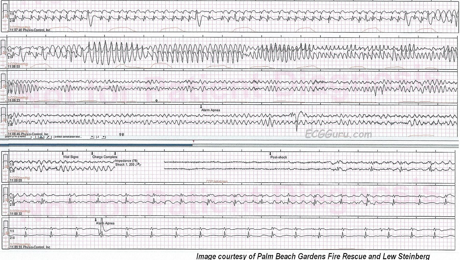
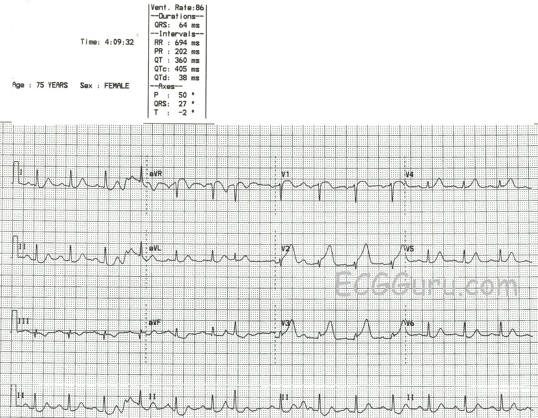
A series of ECGs can be a valuable addition to any teacher's collection.  This series follows a 75-year-old woman through three days, during which she experienced an acute anterior wall M.I., a catheterization with angioplasty and stents placement.

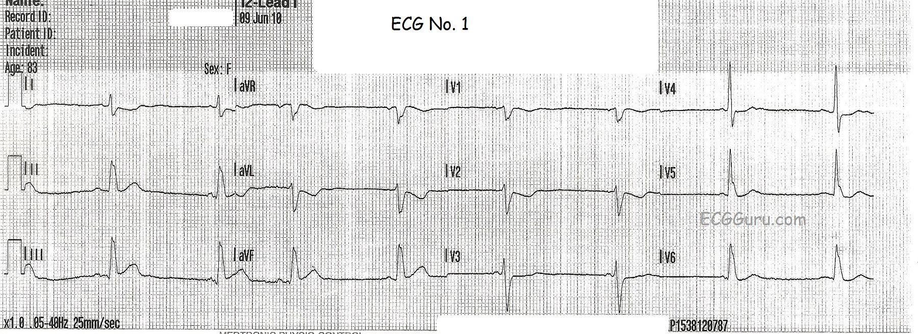
In the first ECG, taken at 4:09 am, the patient has presented to the Emergency Dept. with a complaint of chest pain. (Other details are no longer available).  Although there is some baseline artifact, it appears that the rhythm is sinus rhythm with one PAC (7th beat).  There is subtle but measurable ST elevation in V1, V2, and V3 (anterior-septal leads).  The shape of the STE in V1 is noticeably coved upward.  Even aVR has some STE, with coving.  There is equally subtle ST depression in Leads II, III, and aVF (inferior leads).  Fortunately, there are no pathological Q waves at this point, which would be an indication of necrotic tissue in the area of the M.I. (anterior-septal wall).

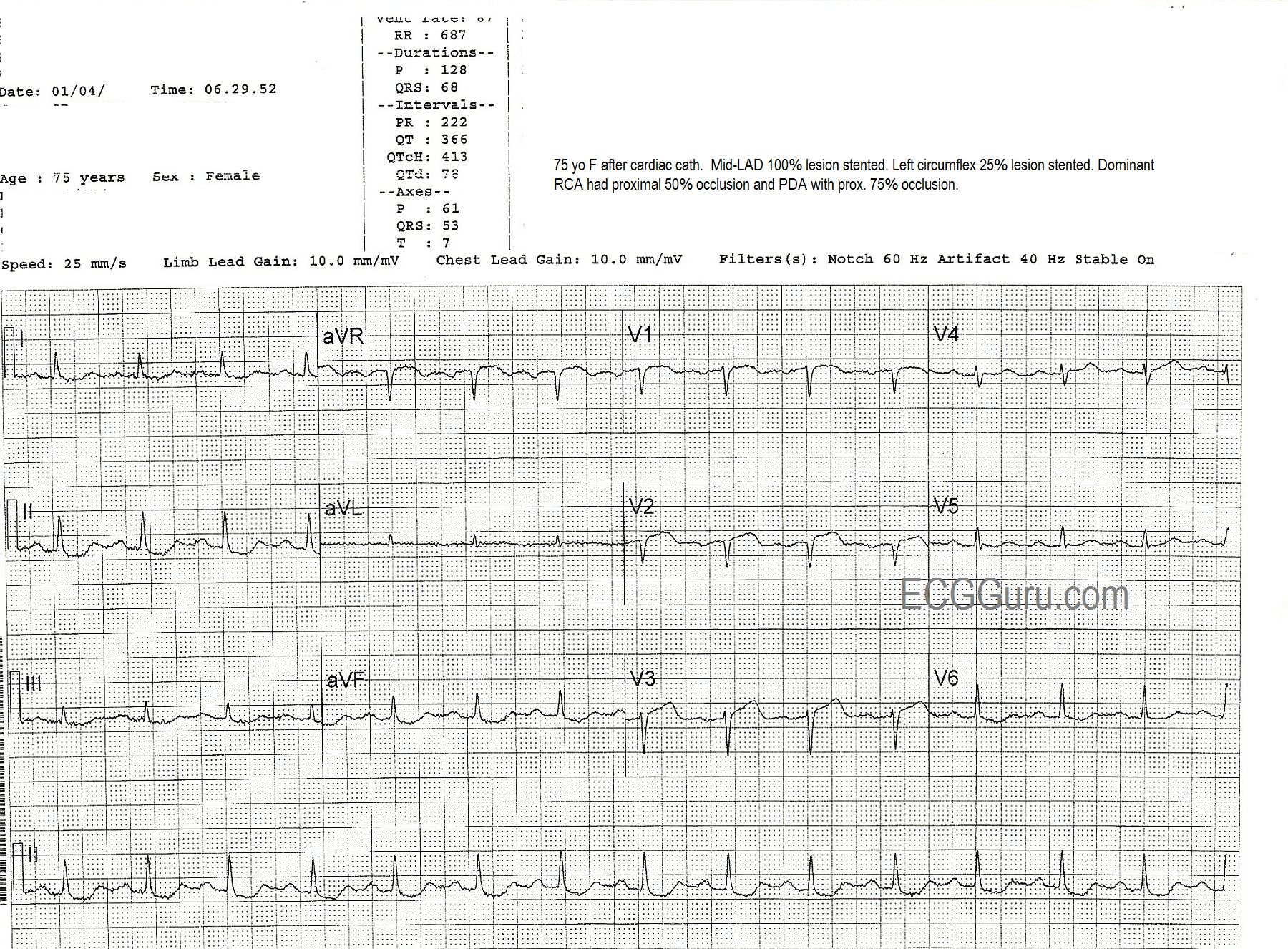
The patient was taken to the cath lab, where it was found that she had a 100% mid-left anterior descending artery occlusion, which was opened and stented.  She also was found to have widespread coronary artery disease, with the left circumflex artery 25% occluded (stented), the right coronary artery (which was dominant) proximately occluded 50% and stented, and the posterior descending artery 75% occluded (stented).

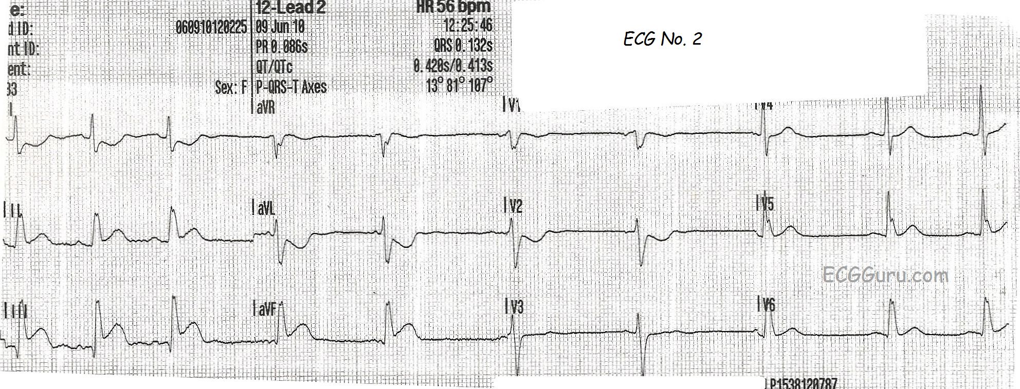
The second ECG, taken at 6:29 the same morning, after the cath procedure, shows some ST elevation with coving remaining in V1 through V3, and also aVR, but now with the loss of R waves in V1 and V2 and loss of R wave voltage in V3.  This represents the formation of pathological Q waves, and can be a permanent change in many cases.

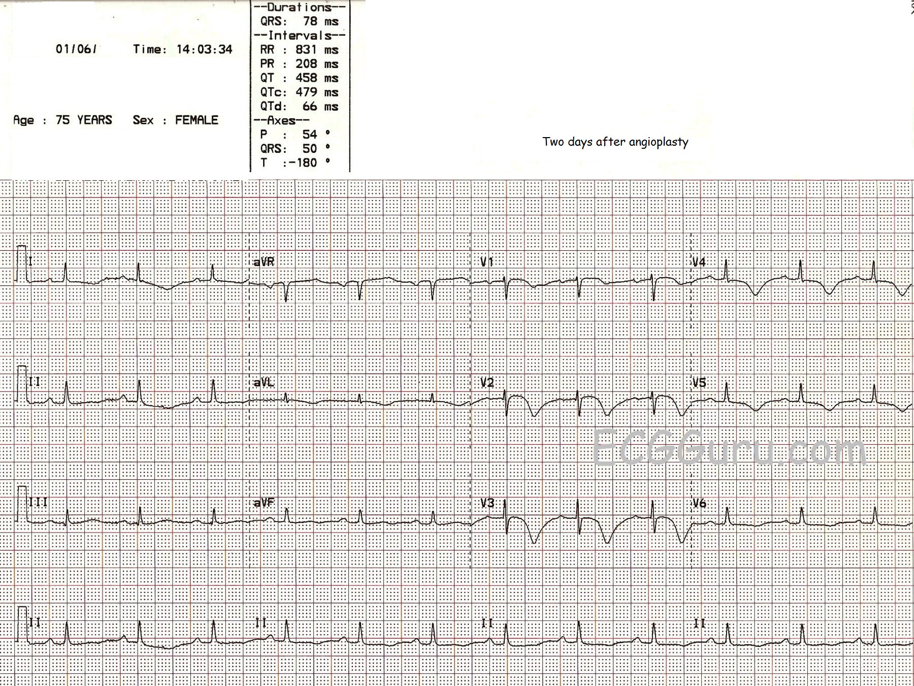
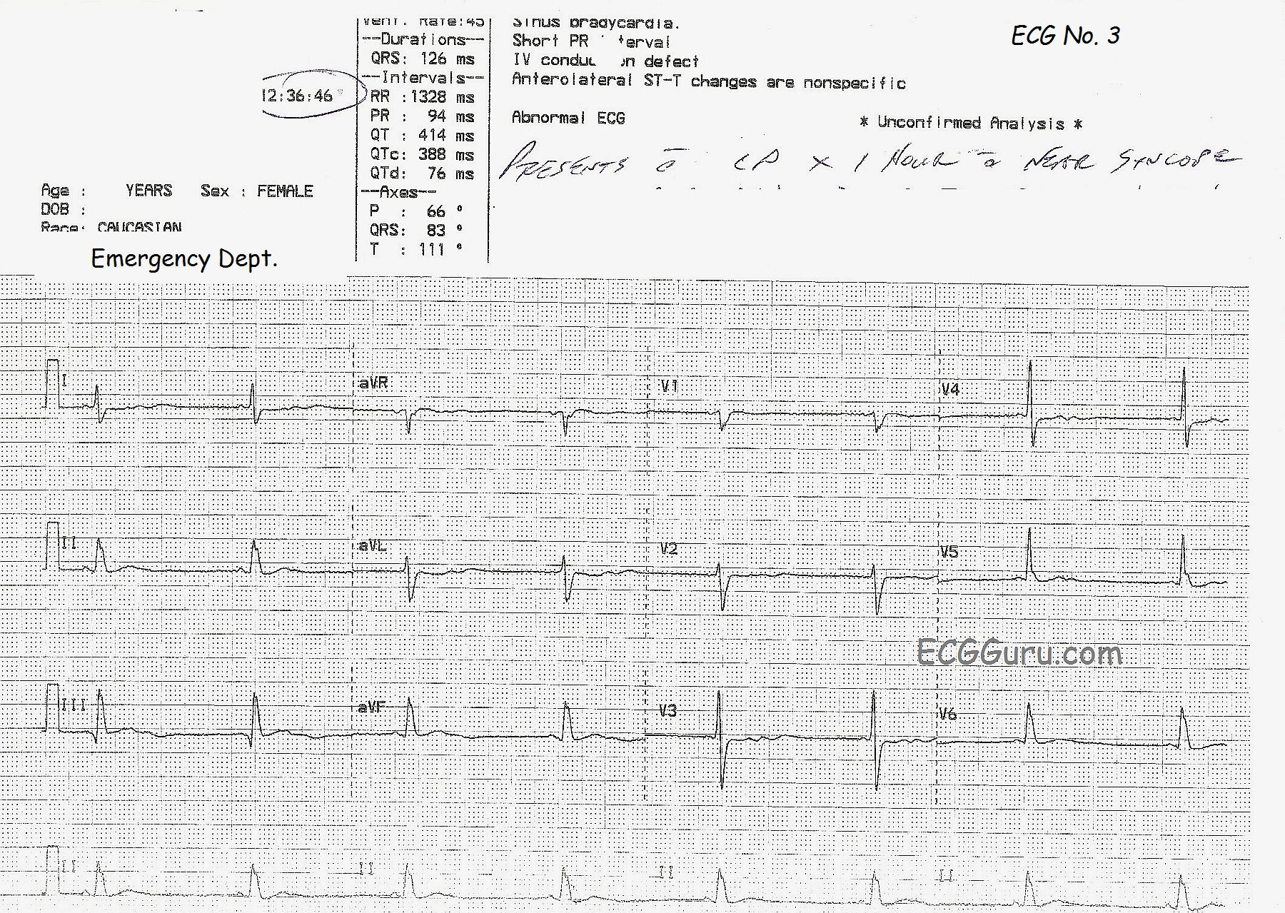
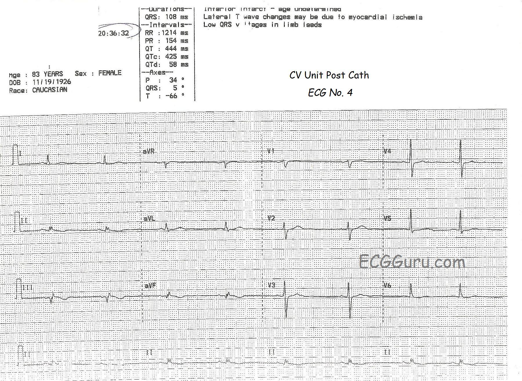
The third ECG, taken two days later in the cardiac step-down unit, shows improvement, and progression toward healing.  The ST segments are still shaped in a slightly coved-upward shape, but they are less elevated.  The R waves have returned.  The T waves in V1-V3 are inverted.  The deeply inverted T waves of V2 and V3, especially, and classic for ischemia, and we even see the "ischemic zone" extending across the anterior-lateral wall, including V4 through V6 and Leads I and aVL.

The patient did very well to discharge, and we don't have followup after that.


Dawn's picture

ECG Basics: Paroxysmal Supraventricular Tachycardia Treated With Adenosine

This series of ECG rhythm strips shows a paroxysmal supraventricular tachycardia successfully treated with adenosine.  The patient was complaining of a rapid heart rate and palpitations, but was hemodynamically stable.  It is not known whether any parasympathetic stimulation, such as a Valsalva maneuver or carotid sinus massage, was used initially.   

The first rhythm strip shows a PSVT, presumably AV nodal reentrant tachycardia, at a rate of about 215 per minute.  (We originally indicated a rate of 240 per minute, but this was a typo). Using the simplist method of determining rate, the six-second method, we see 21, but almost 22, QRS complexes in six seconds. Differential diagnosis would include sinus tachycardia, but this rate is too fast for sinus tach, especially in a resting patient.  Also, sinus tach would slow down as the patient is rested or made more comfortable, and this rate did not vary.  Also, when confronted with a supraventricular tachycardia, one should also consider atrial flutter and atrial fibrillation.  This is somewhat slow for atrial flutter with 1:1 conduction, and that rhythm is much more rare than AVNRT. It is too regular for atrial fibrillation.  So, we are left with the probable diagnosis of paroxysmal supraventricular tachycardia.  The “paroxysmal” part is presumed since AVNRT has an abrupt onset, and the patient’s symptoms started suddenly. 

The second rhythm strip shows what happened after adenosine was administered.  The patient received first a 6 mg dose, rapid IV push.  When that was not effective, he received 12 mg rapid IV push.  The rhythm strip is typical of the first minute or so after adenosine administration.  Adenosine can cause transient AV blocks, escape rhythms, and ectopic irritability.  The half-life of adenosine is only 6 seconds, so the dysrhythmias and uncomfortable symptoms are short-lived.  In this strip, we see frequent PVCs and runs of V tach.  

Dawn's picture

Teaching Series: Atrial Fibrillation With Left Bundle Branch Block

This is a good example of atrial fibrillation with left bundle branch block.  You get two ECGs with this one, because the patient presented to EMS with a fast heart rate, and the rate was slowed with the drug diltiazem.  We do not have any other patient information, unfortunately.

In the first strip, we see a wide-complex tachycardia.  In an emergency situation, with an unstable patient, this rhythm could safely be treated using an emergency ventricular tachycardia (VT) protocol.  In fact, all WCTs should be considered to be VT until proven otherwise.  In most emergency settings, the unstable VT patient would be electrically cardioverted, which will often convert atrial fibrillation as well.  The stable patient with this rhythm would be treated with an antiarrhythmic drug, such as amiodarone.  This may convert or slow down atrial fib.  So, in the initial stages of treatment, differentiating between VT and A Fib is not the first priority.  Assessing the patient's hemodynamic stability and addressing the rate if necessary are the priorities.  

So, how do we know this is NOT VT?  It can be difficult, but in this case, the rhythm, even though fast, is very irregular.   VT is not always perfectly regular, but this irregularly-irregular rhythm points to atrial fibrillation. Also, the pattern of the QRS morphology fits with LBBB.  The criteria for LBBB are:  1) supraventricular rhythm, 2) wide QRS, 3) negative QRS in V1 with positive QRS in Leads I and V6.  If we assume the rhythm is atrial fibrillation, we meet the first criteria.  The other two are self-evident.

After the medication is administered, 10 minutes later, we see the rate slow down.  There is no change in the irregularly-irregular rhythm, and the LBBB pattern remains.  All that has changed is the rate and, hopefully, the patient's symptoms.  This confirms that the original rhythm was not VT.  

Remember, atrial fib lowers cardiac output because there is no P wave - no "atrial kick".  Also, the fast rate associated with new-onset atrial fib often compromises ventricular filling and cardiac output.   LBBB also has a deleterious effect on cardiac output.  Wide QRS complexes indicate that the ventricles are not contracting efficiently and synchronously.  The left ventricle is depolarizing by way of a slow wave of depolarization, rather than all the cells getting the message to depolarize at the same time.  Having these two conditions at the same time can have a very negative effect on cardiac output, leading to CHF.  The first step in treatment often involves simply slowing the rate to normal, which allows for better ventricular filling and decreases the workload on the heart.  Then, the fibrillation and bundle branch block can be addressed.

Dawn's picture

Teaching Tips: ECG Series WO 118: STEMI With Changing ST Elevations

Continuing with our theme of using  series of ECGs for teaching, we present the case of an 83-year-old woman with weakness, chest pain, and near-syncope.  The first ECG, taken by paramedics at her home, shows a sinus rhythm with a slightly wide QRS complex.  At this point, it is unclear whether this represents left bundle branch block or a non-specific interventricular conduction delay. There is the most subtle ST elevation in Leads III and aVF. In LBBB without acute M.I., we would expect the ST segments to be discordant, or opposite, the QRS complexes.   Even more informative is the reciprocal ST depression in Leads I and aVL (and a little in V2).  This can be a sign of inferior wall injury that shows up earlier than ST elevation in the inferior leads.

ECG number 2 was taken during transport to the hospital.  The chest pain continues, and now the ST elevation in the inferior leads is pronounced, and the reciprocal depression in I, aVL, and V2 has also become much more obvious.  V1 would also normally show this depression - a sign of the injury traveling up the back of the inferior wall (posterior).  But if the right ventricle is injured, V1 will have ST elevation.    V1 in this case is probably reflecting the depression from the posterior wall AND the elevation from the right ventricle.  The ST elevation is now prominent enough that the paramedics notified the hospital of a "STEMI Alert".  The QRS remains widened at .13 sec., but the ST elevations and depressions are not opposite the QRS direction, which would be typical for LBBB without an M.I.  Therefore, even if LBBB is present, these ST elevations and depressions would be considered a sign of STEMI.  The rate is slowing in this ECG.

ECG number 3 was taken in the Emergency Department.  The patient's symptoms continued.  The bradycardia is still present, as is the QRS widening.  Now, something new has shown up:  prominent U waves in the precordial leads.  There appears to be T-U fusion.  We do not know the patient's medications or lab results, so the most likely cause for the prominent U waves is the bradycardia.  She does not have other ECG signs of hypokalemia, which is one of the many causes of U waves. 

All our content is FREE & COPYRIGHT FREE for non-commercial use

Please be courteous and leave any watermark or author attribution on content you reproduce.