Submitted by Dr A Röschl on Sat, 05/27/2023 - 01:46
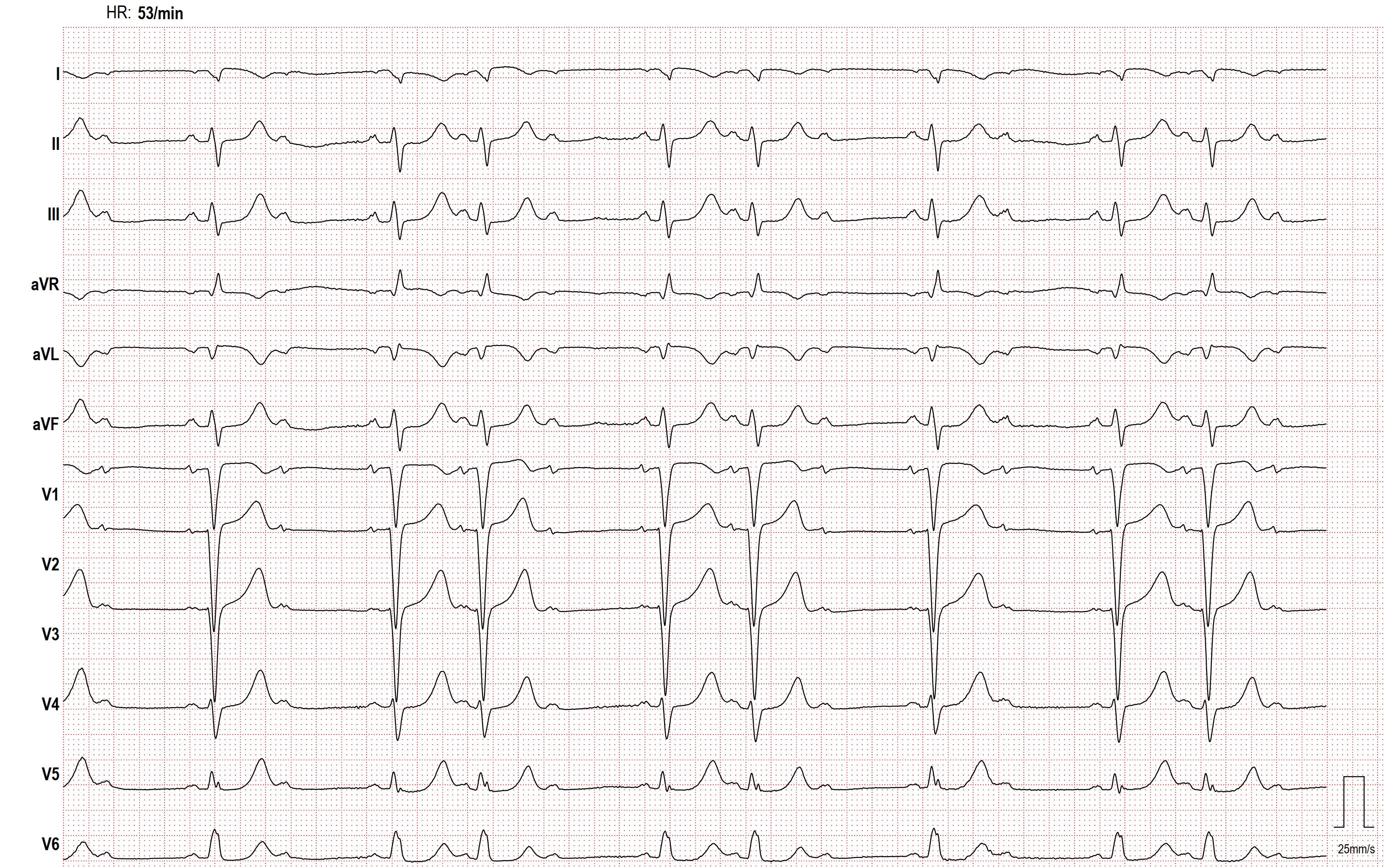
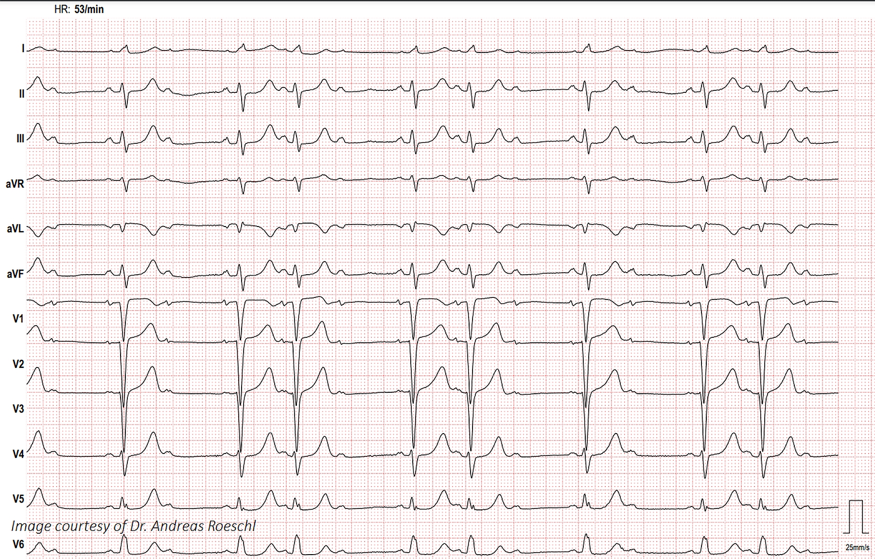
This ECG shows second-degree AV block, Mobitz Type II and an interventricular conduction delay, probably left bundle branch block. The QRS width is about 130 ms, or .13 seconds.
The Patient:This ECG was taken from a 73-year-old man with a history of heart failure with preserved ejection fraction, severe left ventricular hypertrophy, Type II diabetes, and stage 4 chronic kidney disease.He also suffered deep vein thrombosis and is on anticoagulation.He has a recent diagnosis of IgA myeloma.He presented with a complaint of nausea and vomiting and was found to have a worsening of acute kidney infection.There was suspicion of renal and cardiac amyloidosis, but the patient refused biopsy to confirm this.He was started on chemotherapy for multiple myeloma and will be followed as an outpatient.
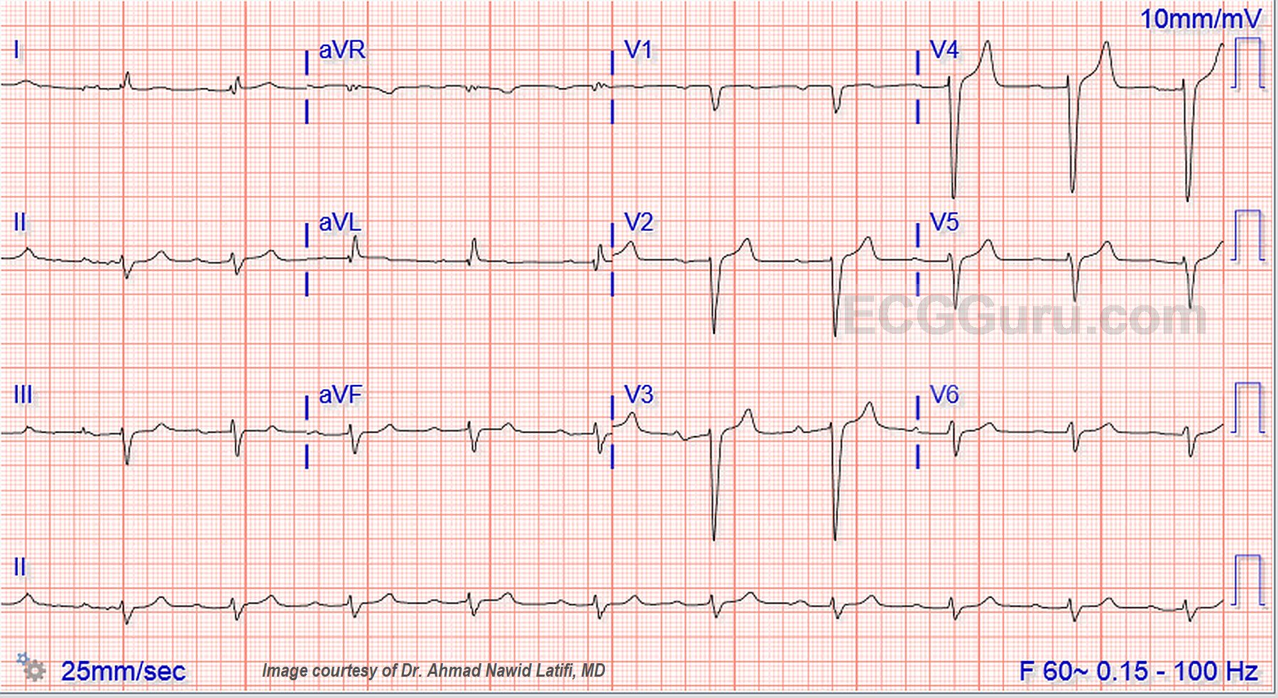
The ECG:The rhythm is sinus at around 60 bpm, although the rate varies a little at the beginning of the strip.The QRS complex is wide at .12 seconds, or 120 ms., representing interventricular conductiondelay (IVCD).The PR interval is .32 seconds, or 320 ms. This constitutes first-degree AV block.There is left axis deviation in the frontal plane and poor R wave progression in the horizontal plane.