This patient is a 50-year-old man with a history of epilepsy and early dementia. He had a VP shunt placed in the hospital and was then discharged home. He became extremely weak, which was not characteristic of him, and 911 was called. He was transported to the hospital uneventfully. He was found to be afebrile.
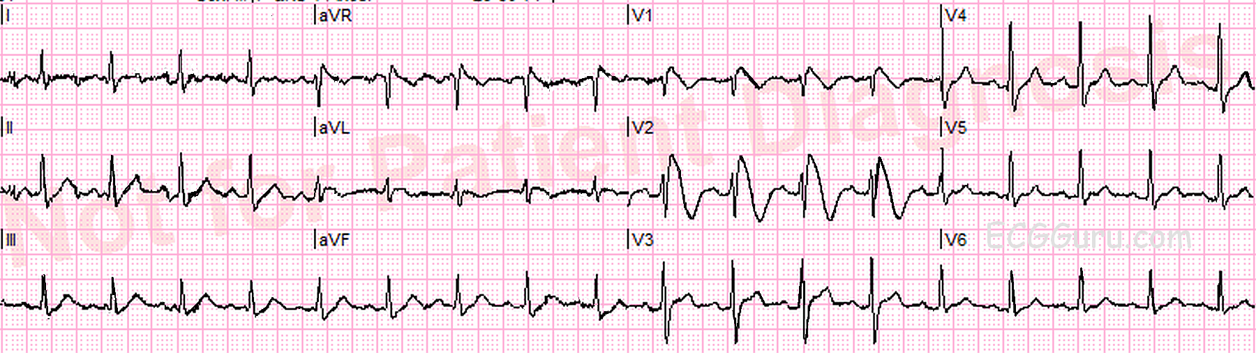
This ECG shows a “classic” Brugada pattern. Brugada Syndrome is a hereditary disease that is associated with a high risk of sudden cardiac death. It is due to a mutation in the sodium channel gene (channelopathy). The ECG characteristics are:
·ST elevation in V1 through V3 of at least 2 mm at the J point in the right precordial leads (V1-V3).
·Coved upward ST segments with negative T waves in the affected leads.
Brugada Syndrome is diagnosed when the ECG pattern exists with one of the following: