Displaying 1 - 4 of 4

Normal Conduction Compared To Conduction Over An Accessory Pathway

Click to open: 
Normal Conduction Compared To Conduction Over An Accessory Pathway

This is an original illustration by Dawn Altman. You may use it free of charge to enhance your presentations or student handouts. Click on image, or right click and SAVE image.

Dawn's picture

Supraventricular Tachycardia in Pediatric Patient With Wolff-Parkinson-White Syndrome

This ECG was obtained from a 13-year-old boy who had previously been in NSR.  At that time, he was diagnosed with Wolff-Parkinson-White syndrome, and he had delta waves.  He now presents with a supraventricular tachycardia.  People with W-P-W have one or more accessory pathways which allow atrial impulses to bypass the AV node and “pre-excite” the ventricles.  An accessory pathway, along with the normal pathway through the AV node, can form a functional circuit, allowing conduction to proceed down one pathway and return up the other, in a rapidly repeating circular motion.  This causes paroxysmal supraventricular tachycardia. 


In W-P-W, the most common type of SVT is AV reciprocating tachycardia (AVRT), also called AV reentry tachycardia.  Conduction through the accessory pathway can be forward (anterograde) or backward (retrograde).  The most common type of reentrant conduction in W-P-W is ORTHODROMIC, which we see here.  The impulse conducts forward through the AV node, and backward over the accessory pathway.  There is no delta wave, as there is no pre-excitation of the ventricles. 


Features of orthodromic AVRT are:

Fast rate (usually approximately 140-250 beats per minute)    

Narrow QRS complexes (unless BBB or IVCD present)   

P waves retrograde, if seen (negative in II, positive in aVR and V1)  

ST depression and T wave inversion common. 


Dawn's picture

Wolff-Parkinson-White Syndrome

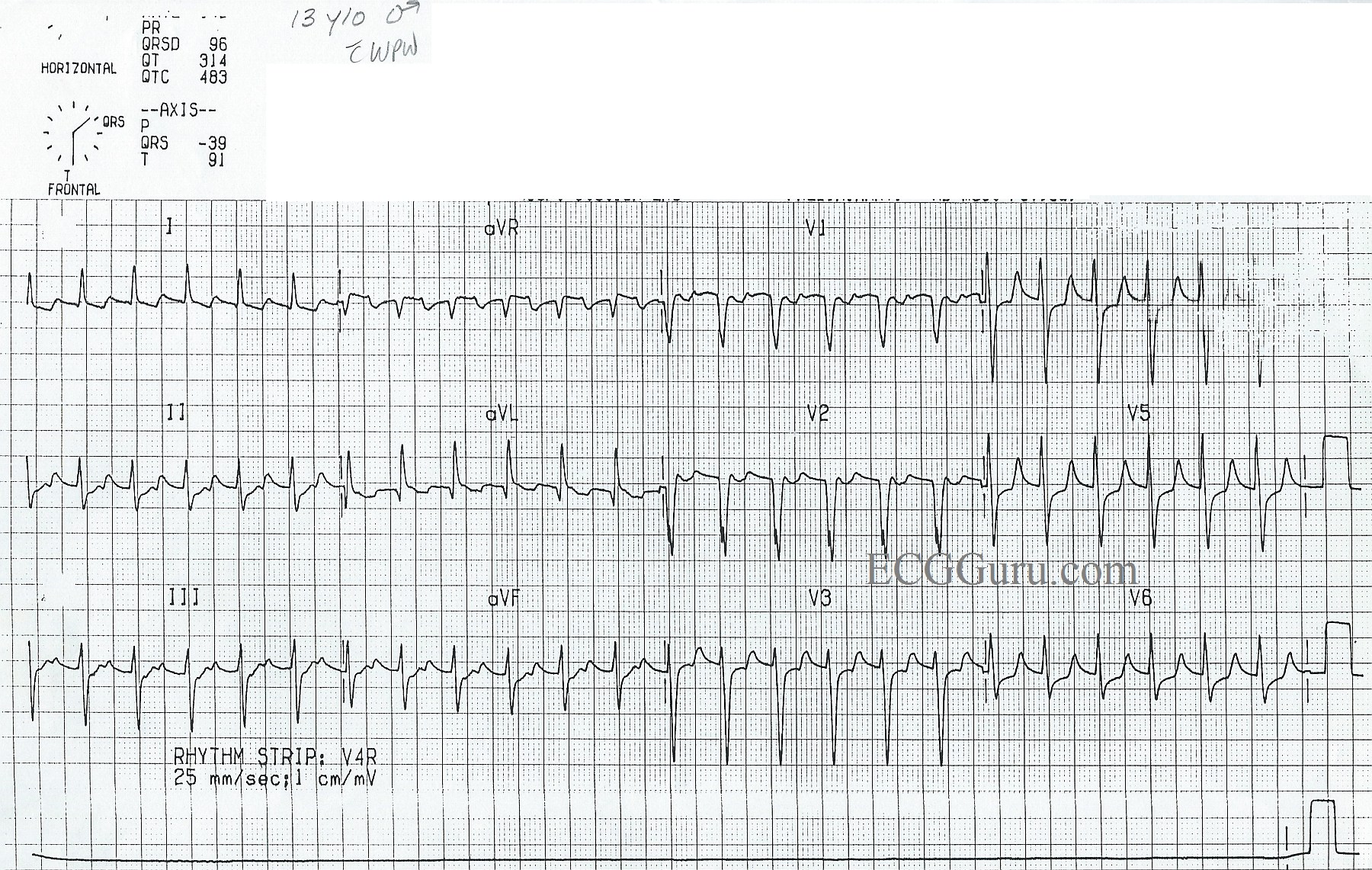
Here is a good example of an ECG showing the signs of Wolff-Parkinson-White syndrome during normal sinus rhythm.  WPW is one of the “pre-excitation” syndromes caused by an accessory pathway between the atria and ventricles. Accessory pathways can bypass the AV node.  These conduction pathways can conduct anterograde (forward toward the ventricles), retrograde (away from the ventricles), most commonly, in both directions.  When the accessory pathway conducts in an anterograde fashion, it causes pre-excitation of the ventricles. See Illustration.

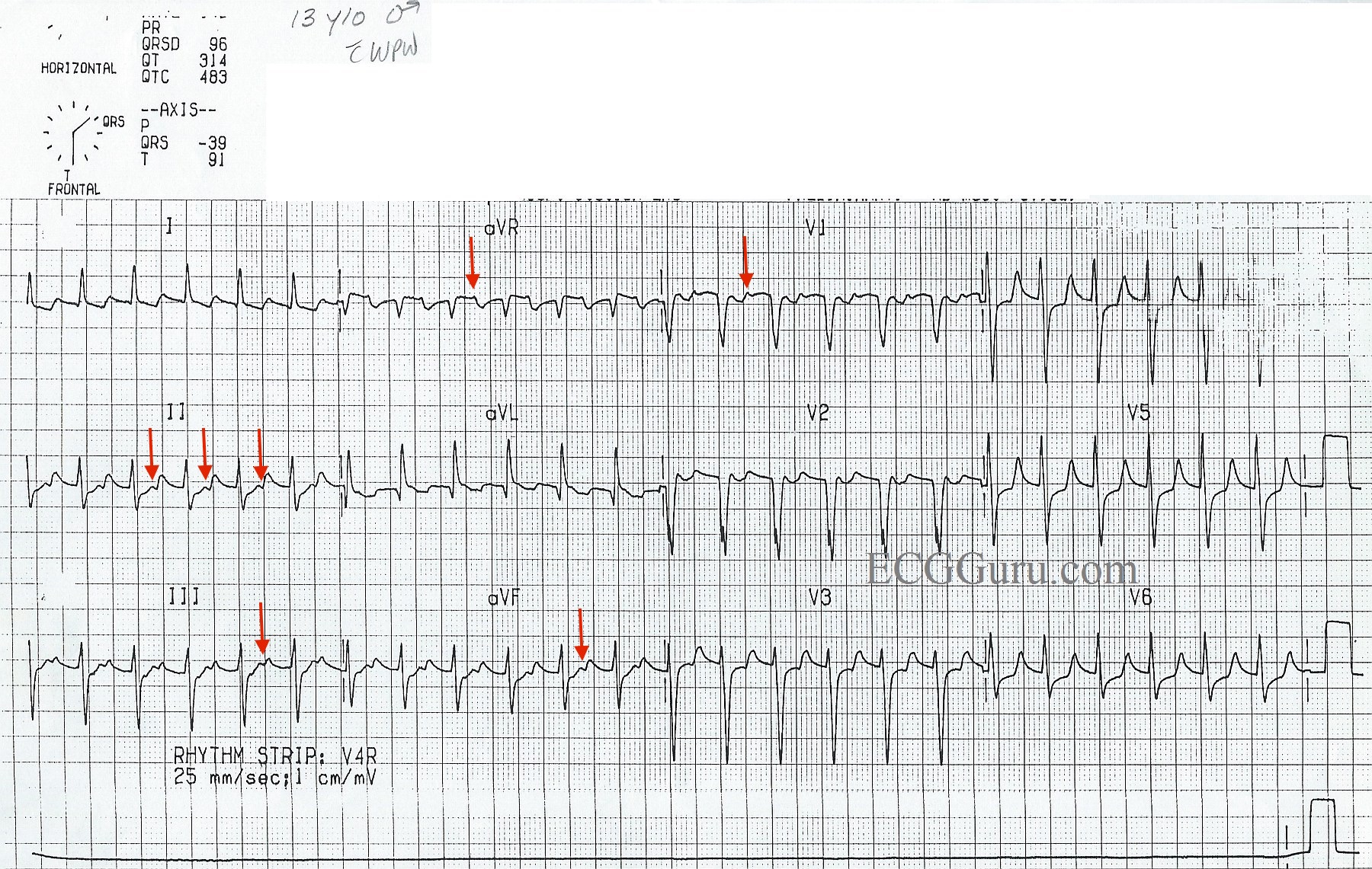
Wolff-Parkinson-White syndrome is caused by an accessory pathway called the Bundle of Kent, which can occur on either the left or the right side of the heart.  The ECG sign of the pre-excitation of the ventricles in WPW is a DELTA WAVE.  The delta wave is seen in normal sinus rates when the accessory pathway conducts anterograde, pre-exciting the ventricles. The delta wave arises at the end of the P wave, eliminating the PR segment in some leads.  In this ECG, the delta waves can best be seen in Leads I, II, aVR, and aVL, as well as in V1, V2, and V3. 

People with WPW are prone to bouts of supraventricular tachycardia, called AV reentrant tachycardia (AVRT).  A circular pathway can form between the normal AV pathways and the accessory pathway, causing an impulse to depolarize the ventricles and return to the atria in a rapidly repeating cycle, causing a paroxysmal supraventricular tachycardia. The delta waves will disappear as they become part of the reentrant cycle.

Tachyarrhythmias can also form when impulses from atrial flutter or atrial fibrillation rapidly enter the ventricles through the accessory pathway without the filtering effect of the AV node.  The extremely rapid rates produced in these situations can quickly lead to deterioration and ventricular fibrillation.


For more information on WPW ECG signs, we recommend Life In The Fast Lane

Dawn's picture

Wolff-Parkinson-White Syndrome in a Four-Year-Old Girl

WPW is one of the "pre-excitation" syndromes caused by an accessory pathway that bypasses the AV node. This pathway is called the Bundle of Kent. This ECG clearly demonstrates the "delta wave" caused by the atrial impulse traveling around the AV node and pre-exciting part of the ventricle. These delta waves are best seen in Leads I and V4 - V6. The delta wave in Lead II is a small notch. A short PR interval is also seen. The accessory pathway can, along with the normal pathway through the AV node, form a circle or circuit, making the heart vulnerable to re-entrant tachycardia. This ECG is from a four-year-old girl who had not been previously diagnosed.

All our content is FREE & COPYRIGHT FREE for non-commercial use

Please be courteous and leave any watermark or author attribution on content you reproduce.