Just like other subjects we are taught in school, ECG interpretation is usually taught in a very basic, simplistic way. As we add to our knowledge, we are able to determine the mechanisms of more complex rhythms.
When I took my first basic ECG rhythm monitoring course, I memorized all the “rules”, and at the end of the course, I thought I could read ANY strip correctly. Then, in real life, I found that some rhythms can’t be interpreted from one lead, or even from one 12-lead ECG.
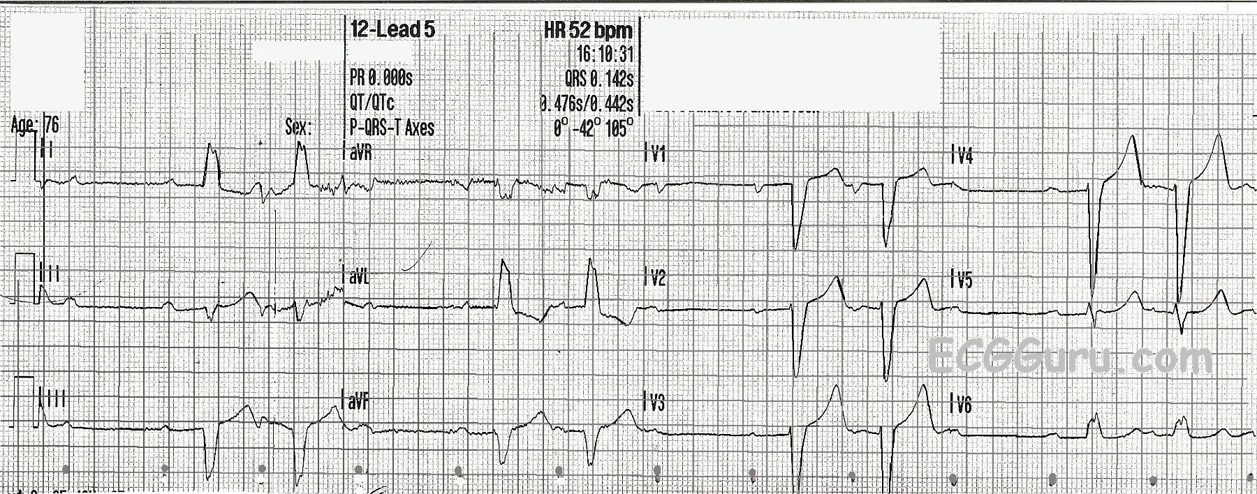
This strip offers advanced readers to challenge themselves, and it offers teachers a chance to show students an “exception to the rules” if it is appropriate for those students. We all learn the classification of second-degree AV blocks: Both Type I and Type II show an underlying sinus rhythm with some P waves conducted and some not. Type I has progressively prolonging PR intervals until a P wave is non-conducted. The cycle restarts after the dropped QRS. Type II has PR intervals that are all the same, and may be prolonged or normal.
In this ECG, you will be able to “march out” a normal sinus rhythm at a rate of 80 bpm. The P waves are marked with small dots at the bottom. Two of every three P waves are followed by QRS complexes. Is it Type I? No – the PR intervals are not prolonging. Is it Type II? The PR intervals are not the same! What is happening?
There is also left bundle branch block, which is a sub-Hisian block. Blocks occurring in the intraventricular conduction system include bundle branch blocks, second-degree AVB Type II, and third-degree AVB with ventricular escape. This group of blocks tends to be more threatening than the blocks that occur in the AV node (second-degree type I and third-degree with junctional escape).