Dawn's picture

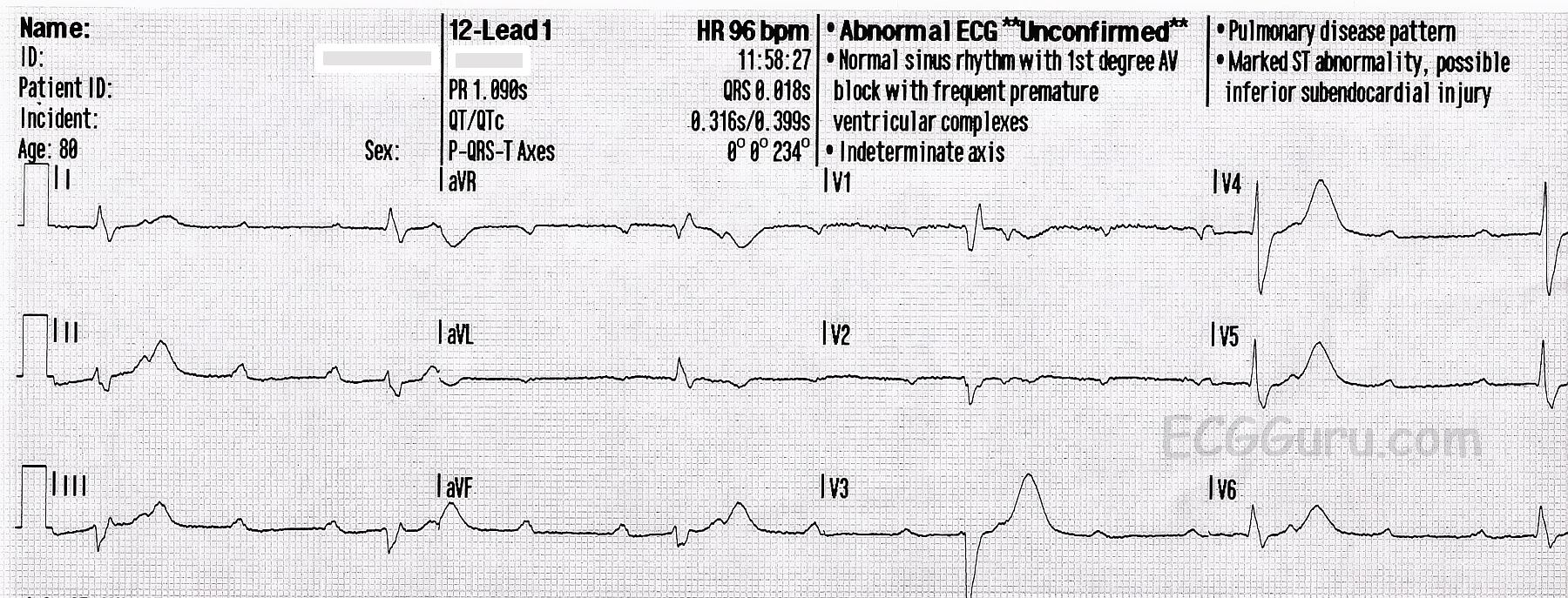
This ECG shows a second-degree AV block, Mobitz Type II.  It is also called “high grade AV block” because there is a 3:1 ratio of P waves to QRS complexes and a resulting slow rate.

Right bundle branch block and left anterior fascicular block are also present, as is common with Type II blocks.  The underlying rhythm is sinus.  Second-degree AVB, Type II, usually represents an intermittent tri-fascicular block:  often right bundle branch block and left anterior fascicular block (hemiblock) are present, and the left posterior fascicle develops an intermittent block.  During times of tri-fascicular block, the P waves are not conducted.  When the posterior fascicle is conducting, a QRS occurs.

A differential diagnosis for this ECG is complete heart block with ventricular escape rhythm.  A longer strip would be needed to see the P waves eventually dissociate from the QRSs, if they are going to do so.  Clinically, there is really little difference in the treatment of a high-grade "second degree" block and a "third degree" block. Both are treated with emergency support of the slow rate, as needed, and then a permanent implanted pacemaker.

It is notable that, in this case, the interpretation given by the machine is completely incorrect, even including the intervals.  This is not common, but does occur.  The machine's interpretation should be considered, but not followed blindly.

Rate this content: 
Average: 4.8 (6 votes)

Comments

ekgpress@mac.com's picture

 
Nice example by Dawn of 2nd-Degree AV Block, Mobitz Type 2. I AGREE with Dawn’s comments. I’ll make a few additional points.
 
It is unfortunate that we do not have any long lead II rhythm strip that is simultaneously recorded with the 12-lead tracing. Making the diagnosis is MUCH easier when you have a long lead rhythm strip …
 
This rhythm should be interpreted systematically — like ANY rhythm. I suggest use of the Ps, Qs, 3R Approach, in which we look for the 5 KEY parameters of rhythm interpretation (in whatever sequence works best for the tracing at hand).
  • P waves ARE present. They are regular.
  • The QRS is wide.
  • The ventricular rhythm is Regular.
  • The ventricular Rate is a bit over 30/minute (though it is difficult to see the grid …).
  • AND — P waves ARE Related to the QRS (note that the PR interval is FIXED preceding each QRS complex). So while I agree with Dawn that ideally we would have a longer period of monitoring — it truly looks as if the PR interval preceding each QRS IS constant, and therefore conducting.
Putting this all together — We have 2nd-Degree AV Block, Mobitz Type II with “high-grade” block ( = 3:1 AV conduction).
 
Now looking at QRS morphology — it is as per Dawn showing RBBB and LAHB. That said — the QRS looks a bit “fragmented” ( = notched) in several leads — which is a sign that prior infarction (ie, scarring) is likely. There is slight hint that the ST segment in lead V1 may be coved and slightly elevated … so it could be that recent infarction is the cause of the AV block — but that is pure speculation (and we don’t know what the “baseline” ECG looks like …).
 
As to the computerized interpretation — I suggest NEVER trusting the computer interpretation for any rhythm other than sinus. There ARE merits to computerized interpretations — but determining complex rhythms is NOT one of them …
 
NICE case!


Ken Grauer, MD  www.kg-ekgpress.com   [email protected] 

All our content is FREE & COPYRIGHT FREE for non-commercial use

Please be courteous and leave any watermark or author attribution on content you reproduce.