Displaying 1 - 9 of 9
jer5150's picture

Jason's Blog: ECG Challenge for the months of July and August, 2014.

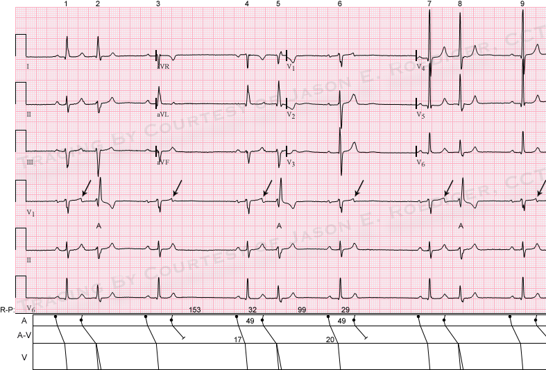
This is an ECG I performed a couple of years ago on an asymptomatic 83-year old man as an outpatient procedure. 

The computer interpreted this as:  "Marked sinus bradycardia [with] Frequent Premature ventricular complexes".  IS THE COMPUTER CORRECT?  Is there more than one plausible interpretation?  What is the differential diagnosis?

jer5150's picture

Jason's Blog: ECG Challenge for the month of December, 2013.

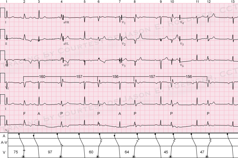
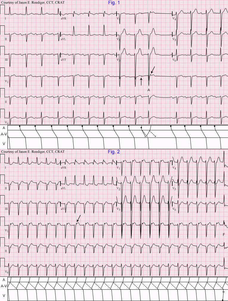
The only patient data I have is that this ECG is from a 73-year old man.  At the request of the site administrator (Dawn Altman), I'm posting this ECG because there isn't one quite like it in the Guru's archives.  Some readers will recognize it as one I recently posted on another website.  This one lives up to the title of "Challenging".  I'll make the same general statement I did on the other website:  You'll need to make careful measurements with calipers on this ECG to come to the correct interpretation.

jer5150's picture

Jason's Blog: ECG Challenge for the month of August, 2013.

Patient of unknown age and gender with a history of atrial fibrillation.  What's your interpretation?

jer5150's picture

Jason's Blog: ECG Challenge of the Week for March 10th - 17th. Mutual mimics

These are two 12-lead ECGs that I performed on two completely different patients several years apart. 

Patient's clinical data (Top ECG):
57-year-old black man

Patient's clinical data (Bottom ECG):
67-year-old black man

Are both of these ECGs showing the same rhythmic pattern?

jer5150's picture

Jason's Blog: ECG Challenge of the Week for Dec. 23rd - 30th.

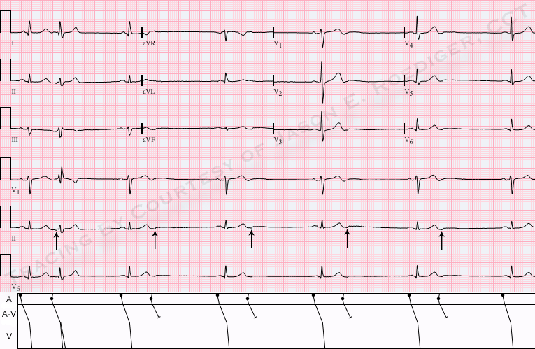
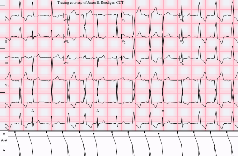
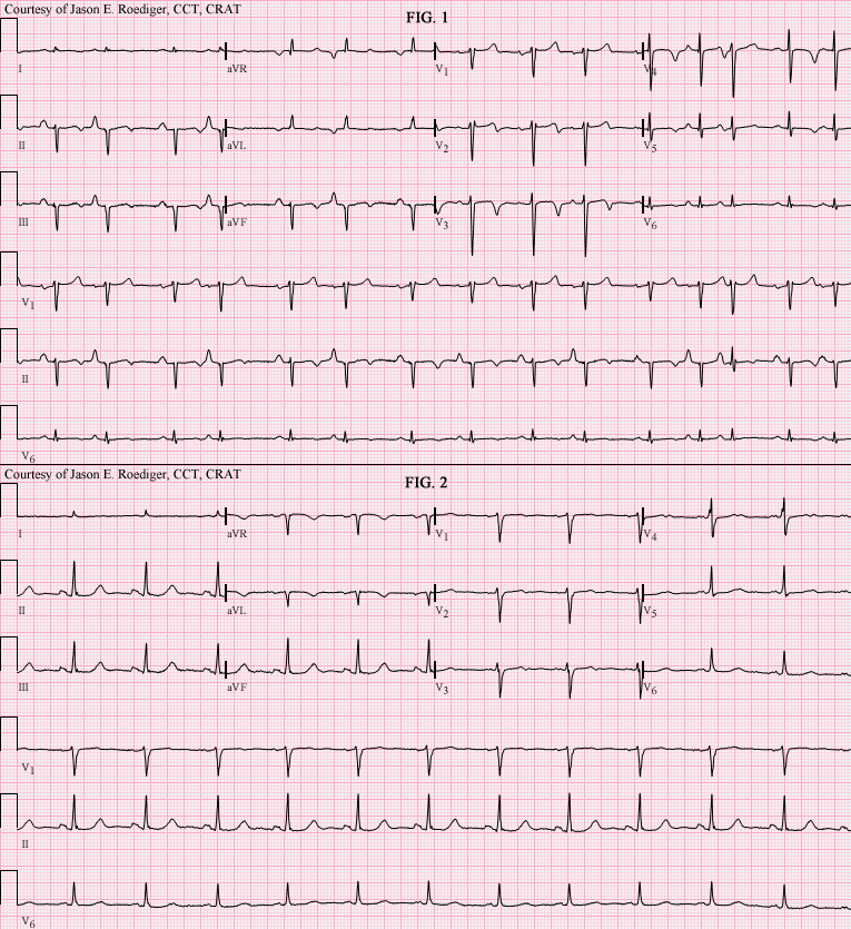
I performed this ECG earlier this year in an outpatient clinic.

Patient's clinical data:  85-year-old white man; asymptomatic. 

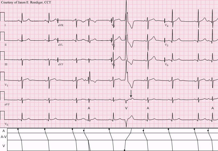
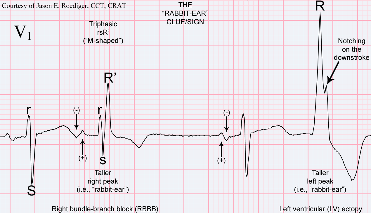
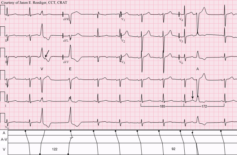
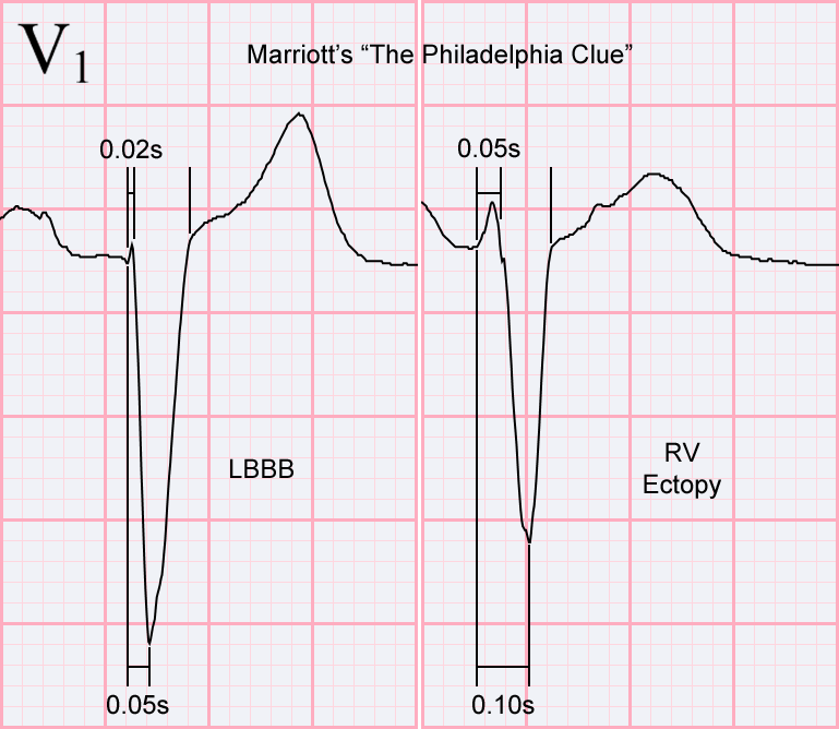
Questions?
(1.)  What rhythm is this tracing showing?
(2.)  What clue/sign is used to differentiate between the two anomalous beats (i.e., 4th and 6th beats).  HINT:  It's named after a part of a certain animal's anatomy.

jer5150's picture

Jason's Blog: ECG Challenge of the Week for Nov. 4th - 18th.

This is a "Routine" resting 12-lead ECG I performed on a patient several months ago in the Outpatient ECG Lab.

PATIENT CLINICAL DATA:
72-year-old white man; asymptomatic; resting comfortably in recumbent position.

This is an exercise in "anomalous" beats.

QUESTION:
(1.)  Specifically, what are 3rd, 4th, and 10th beats?

jer5150's picture

Jason's Blog: ECG Challenge of the Week for August 19-26. The value of a previous, comparative ECG.


Patient clinical data:  68-year-old black man.

Question:
(1.)  What "pseudo" clue in Fig. 1 clinches the source of the mechanism seen in Fig. 2?

jer5150's picture

Jason's Blog: ECG Challenge of the Week for July 8-15. Which lead do both of these ECGs share a “common-thread”?


Two more ECGs classified under the general heading:  “Tracing suggestive of   ____ ”.  I  like ECGs that strongly favor a very specific clinical disorder.

All our content is FREE & COPYRIGHT FREE for non-commercial use

Please be courteous and leave any watermark or author attribution on content you reproduce.