Dawn's picture

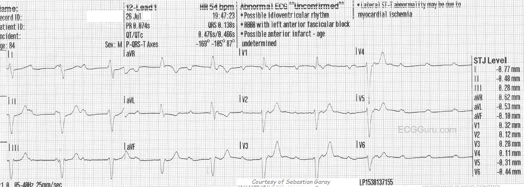
This ECG is from an 84-year-old man who experienced dizziness and a fall.  He was not injured in the fall.  In this ECG, we can clearly see regular P waves at about 110 per minute.  We also see wide QRS complexes at about 52 per minute.  There is AV  dissociation - there are no regular PR intervals, or even progressively-prolonging PR intervals.  The atrial and the ventricles are beating to separate rhythms.  What is interesting about this rhythm is the origin of the escape rhythm.  The wide complex suggests a ventricular focus and the rate suggests supraventricular origin.  Near the end of the ECG, the escape rhythm either fails or slows significantly.  To see the next 12-lead ECG for this patient, go to this LINK.

The second ECG makes it more clear that this is an idioventricular escape rhythm, but the morphology of the QRS complexes suggested that, even in the first ECG when the rate was faster.  There are several clues that this is probably ventricular, including a very "backward" axis with aVR being upright and II, III, and aVF all being negative.  Also, V6 is negative, and there is nearly precordial concordance:  all except V1 are negative.  The morphology of the QRS does not fit a diagnosis of either left bundle branch block OR right bundle branch block.  The evidence points to a ventricular origin for this escape rhythm, and the patient quickly goes on to slow down severely.  Ventricular escape rhythm strongly suggests a sub-Hisian location for the block, and they tend to be more life-threatening than supra-Hisian blocks.

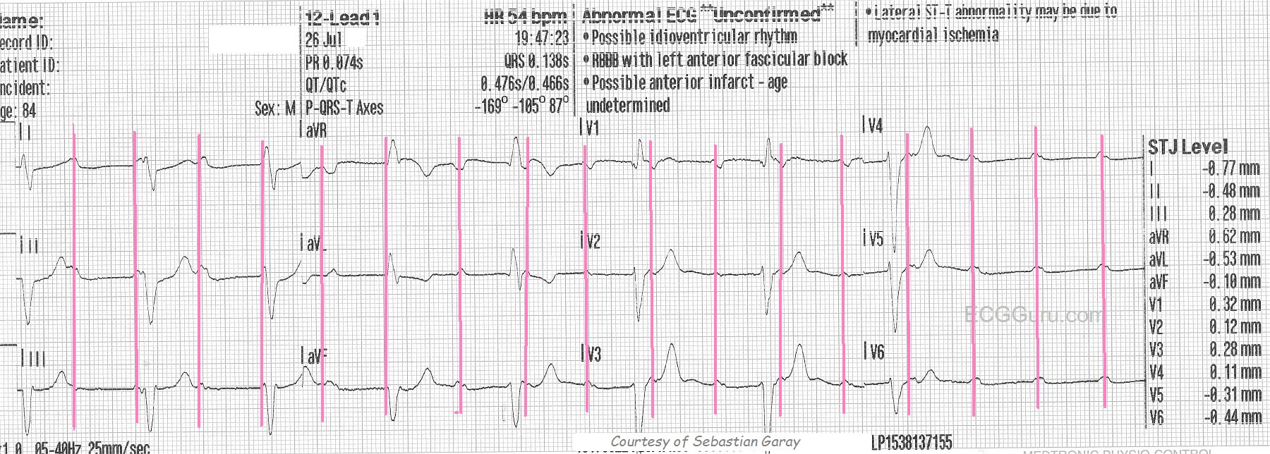
The take-home clinical lesson here is to BE PREPARED for worsening of the rate whenever AV block is present, especially high-grade AV block or sub-Hisian block.  This ECG is a very good one for teaching students to "march out" P waves, and find "hidden" P waves.  We have included a marked copy of this ECG to indicate those P waves.

Thanks to Sebastian Garay for donating these ECGs.


Rate this content: 
Average: 3.5 (4 votes)

Comments

ekgpress@mac.com's picture

     Interesting ECG that explains the reasons for dizziness and the fall experienced by this unfortunate 84-year old man. As per Dawn  the diagnosis of at-the-least advanced AV block is obvious. I'll add a few comments.
 
  • I assess AV blocks in the same manner that I assess any other rhythm  by looking for "Ps, Qs & the 3 Rs" = P waves  QRS width  and Rate, Regularity and Relationship (between Ps & the QRS). This rhythm is no different.
  • The atrial rate is regular at ~ 110/minute (as per Dawn  with red vertical lines highlighting P waves in the lower tracing).
  • The QRS is wide. Ventricular etiology should be assumed unless proven otherwise. QRS morphology totally supports this  as the qR in V1 is not a typical RBBB morphology (ie, no rSR' with taller right rabbit ear the axis is unusual and indeterminate  and the QRS is virtually al negative in V6. This is simply AIVR (Accelerated IdioVentricular Rhythm) = a slightly faster-than-anticipated ventricular escape reason.
  • The initial portion of this 12-lead looks to be obvious 3rd degree AV block (up to beat #6)  as there is complete AV dissociation up to this point with P waves having no relationship to the QRS. Most of the time with 3rd degree  the ventricular rate is regular (or at least fairly regular and that is what we see for these first 6 beats with certain P waves failing to conduct despite seemingly having adequate opportunity to do.
  • Things academically become a little less clear after the 6th beat  as it looks like the PR interval MIGHT be the same for beats #6,7,8 .... Making it more difficult to tell is the unfortunate lack of a long lead II rhythm strip  so I'm just not sure if the PR interval is or is not the same for these 3 beats. If it was  then we have Mobitz II rather than 3rd degree AV block toward the end of this tracing (which finishes with 4 consecutive nonconducted P waves). If on the other hand, the PR interval does continue to change some  then we just have a period of ventricular standstill (that we don't know for how long this lasts given termination of the tracing with a series of nonconducted P waves). Clinically  it does NOT matter of course since the "theme" is at-the-very-least high-grade AV block that is symptomatic and requires pacing.
  • MY GUESS  given the link that Dawn provides to the follow-up tracing  is that there was ventricular standstill with disappearance of this accelerated ventricular pacemaker. NOTE how QRS morphology in lead I especially of the follow-up tracing is different  suggesting there is now a slower ventricular escape focus that has replaced the AIVR focus that failed. Hopefully a pacer was placed before this 2nd focus also failed ....

NOTE: We review the Basics of AV Block in a 50-minute Video (GO TO www.avblockecg.com ).

  • CLICK HERE — to download a pdf on the Basics of AV Block (excerpted from my ECG-2014-ePub).
  • This case is linked to my ECG Blog #104.

Ken Grauer, MD  www.kg-ekgpress.com   [email protected] 

All our content is FREE & COPYRIGHT FREE for non-commercial use

Please be courteous and leave any watermark or author attribution on content you reproduce.