Dawn's picture

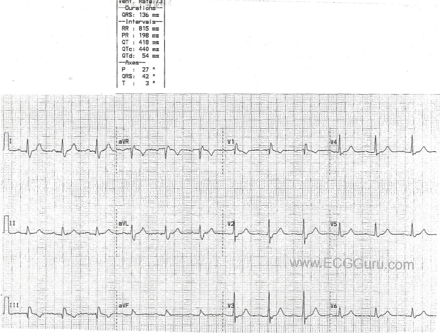
This ECG shows a normal sinus rhythm at 73/min. and a right bundle branch block.  The diagnostic characteristics of RBBB are:  wide QRS (greater than .12 sec.), supraventricular rhythm (in this case NSR), and an rSR' pattern in V1 with a small, wide S wave in Leads I and V6.  The R' and small s waves represent the right ventricle depolarizing slightly AFTER the left ventricle.  In fact, in RBBB, each lead should look pretty normal at first, with a terminal right ventricular depolarization wave added to each QRS complex.  This is because the left ventricle depolalrizes normally.  In this patient, R waves progress fairly soon in the precordial leads (V1 through V2).  It is not known in this case if it is due to poor electrode placement or patient causes.

Rate this content: 
Average: 3.5 (2 votes)

Comments

ekgpress@mac.com's picture

Good teaching example of RBBB posted by Dawn this week. I'd make the following teaching points regarding this tracing and ECG diagnosis of RBBB in general:

  • Diagnosis of RBBB and LBBB can be simplified into a method that allows accurate diagnosis within 2-3 seconds. This is because: i) You only need to look at 3 leads in order to diagnose the type of conduction abnormality = leads I, V1 and V6; and ii) There are only 3 possible answers when you have sinus rhythm with a wide QRS (assuming no WPW) = 1-RBBB; 2- LBBB; and 3- IVCD (IntraVentricular Conduction Defect). While I definitely DO look at all 12 leads for additional clues/findings - you only need 3 leads to determine which of the above 3 conduction blocks is present. For those interested - I've made a web page on the Basics of BBB that reviews full details of this approach, including links to numerous explained BBB examples. 
  • RBBB may be "complete" with a QRS duration of 0.11 seconds. This is different than LBBB - which requires a QRS duration of at least 0.12 seconds to be complete. This makes sense. The RV is not as thick as the LV - so complete RBBB might not result in as much delay as LBBB would.
  • The S waves seen in lateral leads I and V6 are not necessarily small with RBBB. They may actually be quite large. The KEY - is that with typical RBBB - there are wide terminal S waves (that may be shallow or deep). The reason these lateral S waves are wide - is that it takes longer for the impulse to traverse the RV via nonspecialized myocardial fibers (since the RBB is blocked).
  • The expected ST-T wave response to simple complete RBBB is present here. That is - ST-T waves in the 3 KEY leads (I,V1,V6) is oppositely directed to the last QRS deflection. Thus, the T wave is upright as it should be in leads I,V6 (opposite the negative terminal S wave) - and the ST-T wave is negative as it should be in V1 (opposite the positive R' in V1). If ST-T waves deviate from this expected direction - suspect something else (ie, ischemia, infarction) is going on (Full details in the pdfs at the link I give above re Basics of BBB).
  • Q waves are present in lead III and a tiny q in lead II of this tracing. The meaning of this is uncertain - but it might reflect prior inferior MI. Since RBBB is a "terminal delay" - it does not alter the initial vector of ventricular depolarization. As a result - you will usually be able to see infarction Q waves despite RBBB. Given the tiny size of the q in lead II and hte absence of a Q wave in aVF - we can't be certain if the large Q that we do see in lead III indicates prior inferior infarction or is simply an isolated Q that may sometimes be seen in lead III.


Ken Grauer, MD  www.kg-ekgpress.com   [email protected] 

All our content is FREE & COPYRIGHT FREE for non-commercial use

Please be courteous and leave any watermark or author attribution on content you reproduce.