
Submitted by Dawn on Fri, 07/13/2012 - 23:28
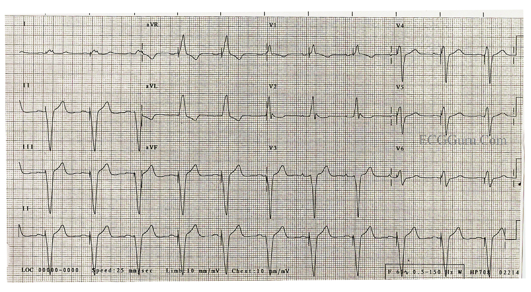
This patient's electronic pacemaker has pacing electrodes in the right atrium and the right ventricle, which is typical. In this ECG, the patient produces P waves, but fails to transmit them to the ventricles due to AV block. The electronic pacemaker SENSES the native P waves, and TRIGGERS the ventricular wire to pace the ventricle in response. Using this pacemaker function, the heart rate can be controlled by the patient's own nervous system, as P waves are produced naturally. Should the patient fail to produce a P wave within rate guidelines set by the implanting physician, the pacer will pace the atrial as well.
The QRS complex is wide when only the right ventricle is paced, as the impulse has to travel from the RV to the LV cell-by-cell. This slows conduction in the same way a ventricular rhythm like VTach is slowed, producing a wide QRS. Remember, a wide QRS causes lower cardiac output, as much as 15%.
For your students who are learning axis, show them an illustration of the heart with the pacing wire in the right ventricle. It is easy to see why the axis is leftward when the left ventricle is being paced from the right ventricle.
Related Terms:
Rate this content:
All our content is FREE & COPYRIGHT FREE for non-commercial use
Please be courteous and leave any watermark or author attribution on content you reproduce.