
Submitted by Dawn on Wed, 04/18/2012 - 11:41
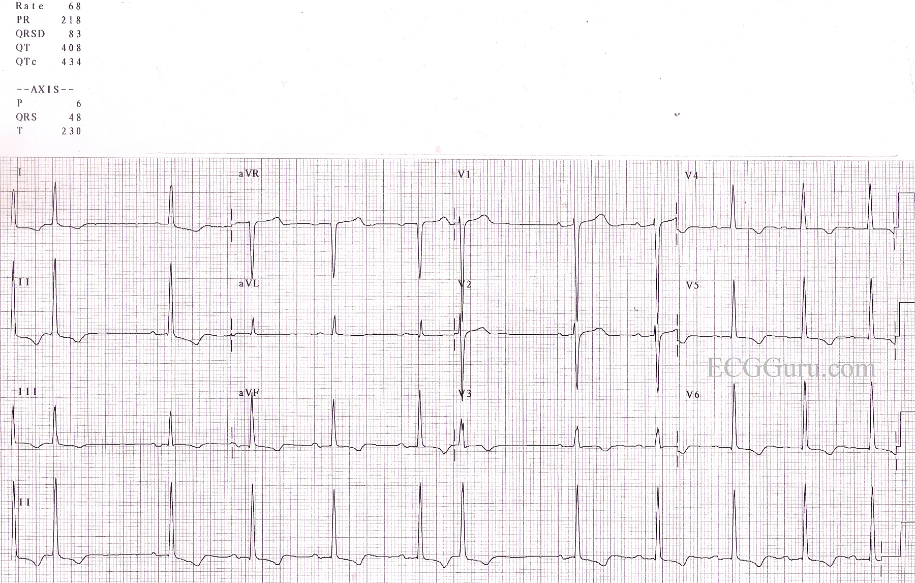
This ECG is from an elderly man with long-standing hypertension. It is a good example of LVH, with tall QRS complexes in the left-sided leads (V5, V6) and deep QRSs in right sided chest leads V1 and V2), but a rather unusual axis, in that it is normal, and we often seen left axis deviation with LVH. His hypertrophy is not severe as seen on imaging studies, but he shows an obvious "strain" pattern of sloping ST depression and T wave inversion in leads with upright QRS complexes. The signs of LVH are subtle, but when viewed as a whole ECG, the pattern seems more obvious. Always consider body shape, also, as a very thin person may produce more voltage on the ECG since the electrodes are close to the heart, and a person with a large chest may seem to minimize the QRSs, as we sometimes see with COPD. For more discussion of LVH criteria, go to the "Favorites" page. Every ECG blogger on the blogs list will have a discussion of LVH criteria and significance.
Related Terms:
Rate this content:
All our content is FREE & COPYRIGHT FREE for non-commercial use
Please be courteous and leave any watermark or author attribution on content you reproduce.