Dawn's picture

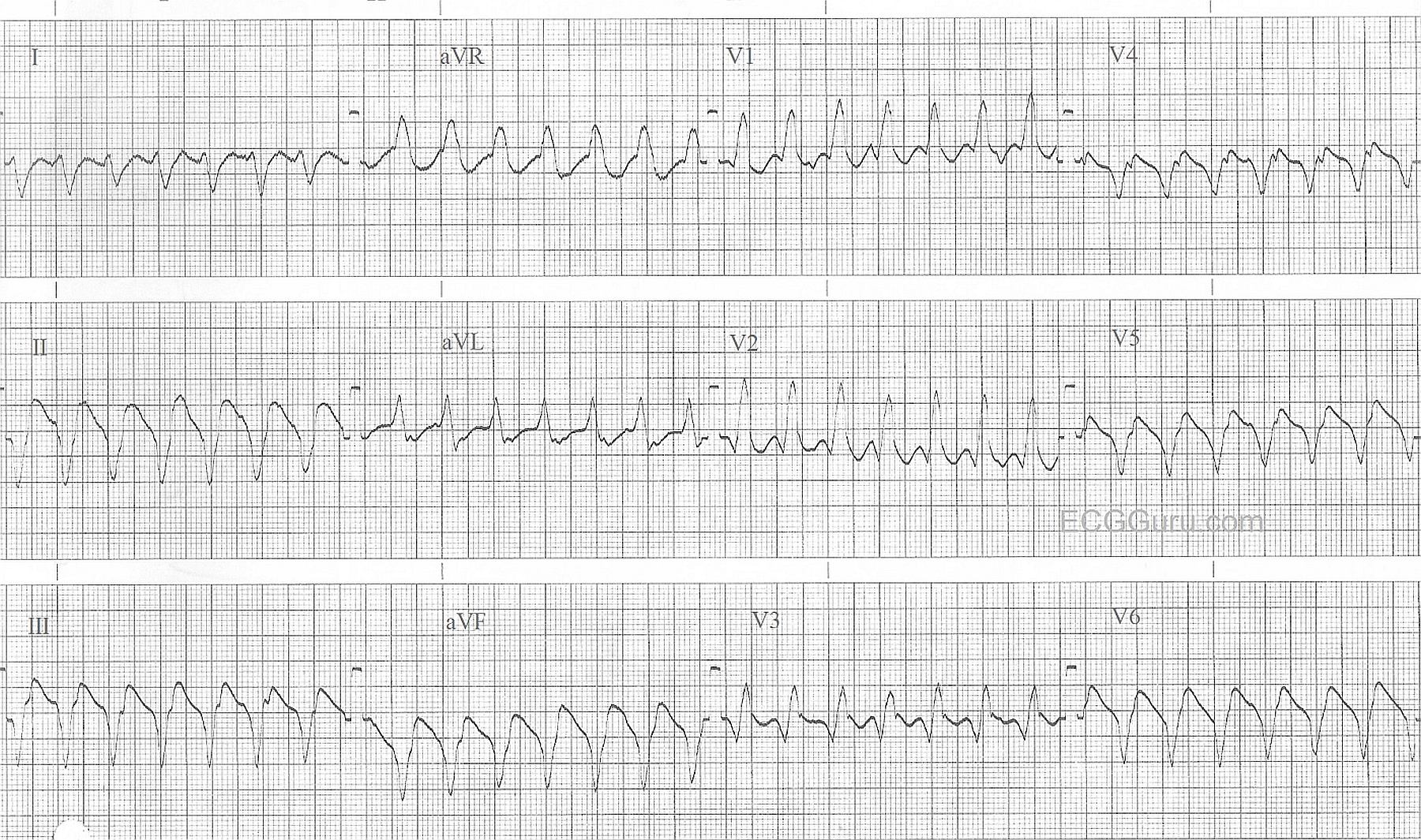
This wide-complex tachycardia is ventricular tachycardia.  Along with the wide QRS and the fast rate, features which favor a diagnosis of VT over BBB include:  backwards (extreme right) QRS axis, negative QRS in V6, and an apparently monophasic QRS in V1, as opposed to the rSR' pattern of right bundle branch block. 
Remember, ALL wide-QRS tachycardias should be treated as V Tach until proven otherwise, as it is a life-threatening arrhythmia.  Factors which lower cardiac output during V Tach include:  Fast rate, wide QRS, and lack of P wave preceding the QRS.  The sudden severe lowering of perfusion that usually accompanies V Tach can lead to rapid deterioraton and ventricular fibrillation.

For discussions by Jason Roediger (ECG GURU extroidonairre) on recognizing ventricular tachycardia, go to this LINK, and this LINK.

Rate this content: 
Average: 4.8 (5 votes)

Comments

ekgpress@mac.com's picture


I would interpret the ECG in the above Figure as follows:

  • The rhythm is a regular WCT ( = Wide-Complex Tachycardia) at a rate of ~ 165/minute. Normal sinus P waves are absent.
  • Several leads seem to have deflections that may reprsent atrial activity (possibly retrograde V-A conduction  if the notching after the QRS in aVL is real). That said  atrial activity is neither clear enough nor consistent enough for me to be comfortable in determining its role, so this is not helpful to me in the diagnosis of this arrhythmia.
  • There is NO History given .... This is important because knowledge that the patient is an adult of a certain age who has underlying heart disease clearly increases statistical likelihood that a WCT is VT (Ventricular Tachycardia).
  • Thus, we are left with the Differential Diagnosis of a Regular WCT Rhythm without normal sinus P waves. One should think of 10 possible entities the first 8 of which are VT, VT, VT, VT, VT, VT, VT and VT. If this patient was an adult with a history of known structural heart disease  then the odds increase to 90+% likelihood of VT even before you look at the tracing itself!
  • While there are numerous criteria to look at in deciding whether a WCT rhythm is VT or SVT (SupraVentricular Tachycardia) with either preexisting Bundle Branch Block or Aberrant Conduction — We favor our "3 Simple Rules". In this case  All 3 of the features we look for are abnormal (even just 1 of these features is enough to greatly increase the odds of VT). That is: i) There is extreme axis deviation (ie, the QRS is all negative in either lead I or lead aVF); ii) The QRS is all negative in lead V6; and iii) The QRS is "ugly" (ie, not resembling any known form of bundle branch block and/or hemiblock).
  • In addition the QRS in lead aVR is all positive. This finding (as well as the finding of an all negative QRS in lead V6 mean that the impulse originates from the apex  and the only thing that does this is VT.
  • The fact that the QRS is all negative in V4,V5,V6 rules out any possibility of WPW (no accessory pathways originate from the apex ... ).
  • BOTTOM LINE: We can confidently say this rhythm is VT.

For full Review of the above Criteria — Please check out my ECG Blog #42 (GO TO  http://tinyurl.com/KG-Blog-42 ).

  • For additional discussion on the differential of the regular WCT — Please check out my Comment on the 6/15/2016 ECG Guru.

Ken Grauer, MD  www.kg-ekgpress.com   [email protected] 

All our content is FREE & COPYRIGHT FREE for non-commercial use

Please be courteous and leave any watermark or author attribution on content you reproduce.