Dawn's picture

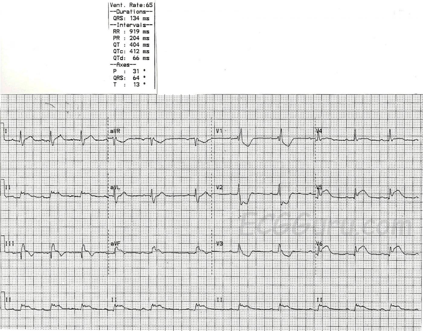
This ECG shows two obvious abnormalities, right bundle branch block AND inferior wall M.I.  It is also a good teaching example of how the terminal wave of RBBB can be mistaken for the ST elevation of M.I.

First, check this ECG to see if it meets the criteria for right bundle branch block:

1)  The QRS will be wide. That is, it will be greater than or equal to .12 seconds (120 ms).  In this case, the QRS is 134 ms.

2)  The rhythm will be supraventricular.  Supraventricular rhythms originate from above the ventricles.  This ECG has P waves before each QRS.  Even though the rhythm is irregular, slowing down during this recorded period, it is a sinus rhythm.

3)  The QRS will have a terminal wave after the "normal" part of the QRS.  This represents the right ventricle depolarizing late.  It is very easily seen in V1, which normally has an rS pattern, and with RBBB has an rSR' pattern, making it appear upright.  V6 and Lead I will show this terminal wave as a wide little s wave.

As mentioned, there is also an acute inferior wall M.I. here.  The ST segment elevation in Leads II, III, and aVF are actually quite subtle.  The flat top of the ST segments gives them away as abnormal, along with the associated ST elevations in V5 and V6, and the reciprocal ST depressions in V1 through V3.  Normally, in IWMI, there will be reciprocal ST depressions in Leads I and aVL, but the elevations they are reflecting are very subtle, and so, therefore, are the depressions. 

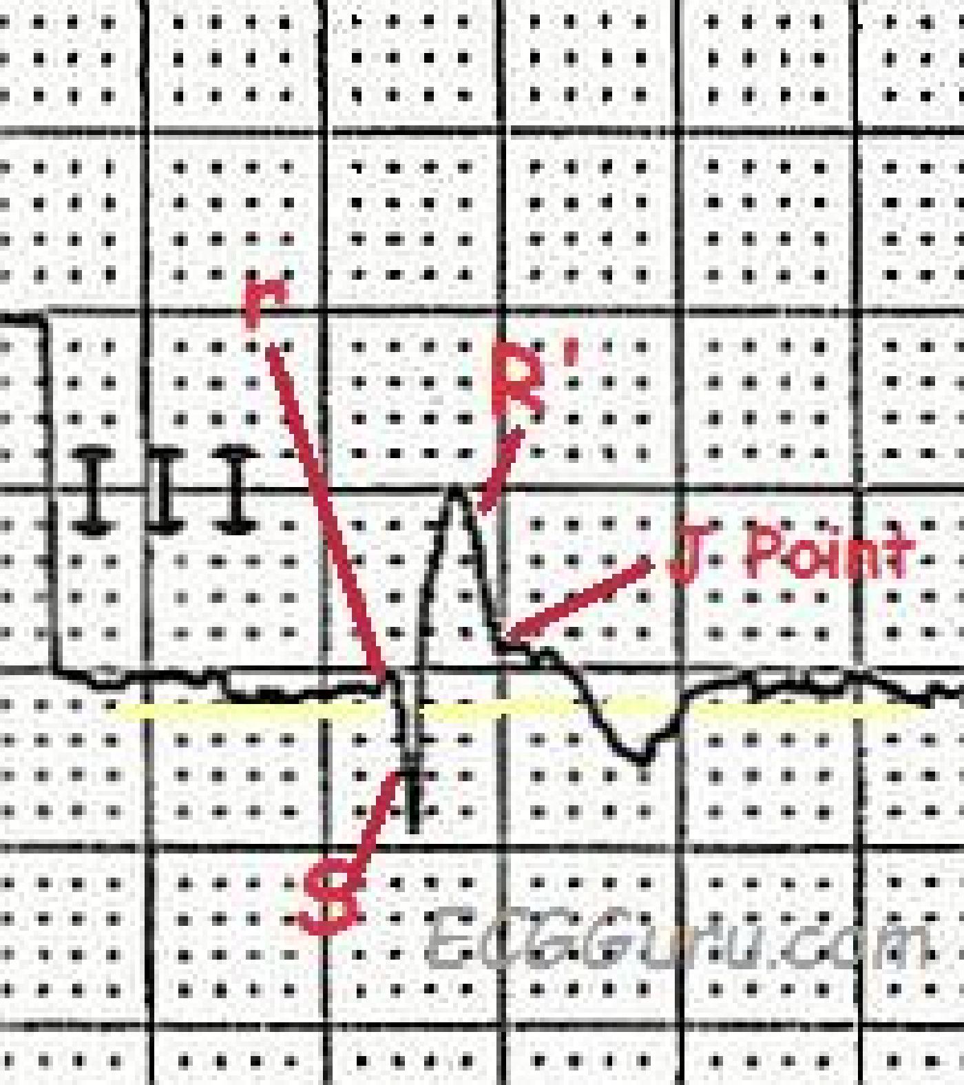
The tricky thing about this ECG is that you must look carefully at the inferior wall leads to see the true ST elevation, which, as mentioned, is subtle.  The RBBB terminal wave of the QRS complexes in Leads III and aVF is upright, and is often mistaken for ST elevation.  Remember, ST segments are smooth from the end of the QRS to the peak of the T wave.  See the detail illustration.

This ECG is suitable for your classes from beginner level (rate variation in sinus rhythm) through advanced (clinical significance of RBBB in acute M.I.).  It also offers an example of reciprocal ST changes, and of a situation where the inferior leads II, III, and aVF are related to the low lateral leads V5 and V6 by a shared blood supply.

Rate this content: 
Average: 4.6 (9 votes)

Comments

ekgpress@mac.com's picture

     
Very interesting tracing - about which I'd make a number of additional points.
  • The rhythm is not regular (See the long lead II at the bottom of the ECG). This is relevant - given the acute MI. That said - despite the baseline "noise" ( = artifact) - I believe the mechanism of the rhythm is sinus - which in this case is consistent with fairly marked sinus arrhythmia. Given acuity of the tracing - this sinus arrhythmia may be part of the overall picture rather than a normal variant.
  • The QRS is wide as Dawn indicates. Although debated - I favor allowance of QRS width to 0.11 second as acceptable for fulfilling criteria for complete RBBB (Right Bundle Branch Block). The normal adult RV (right ventricle) is 1/3 as thick and has ~ 1/10th the mass of the normal LV. As a result - I have never understood why certain experts require equal amount of QRS prolongation to 0.12 second for BBB to be "complete". IF the ECG morphologic criteria for RBBB are typical (ie, rSR' in V1 and wide terminal S waves in both leads I and V6) - then I favor accepting a QRS duration of 0.11 second (rather than 0.12 second) as sufficient for complete RBBB.
  • That said - NOTE that a terminal S wave is not present in lead V6 in this case! This situation is not uncommonly observed when RBBB complicates acute infarction, as is the case here. Whether this is the result of lateral precordial J-point ST elevation impeding formation of a terminal S wave in lead V6 - or simply lack of sufficient time for this terminal S wave to develop - vs other alteration in depolarization orientation resulting from acute infarction is uncertain. The point to emphasize is that in this case despite lack of a terminal wide S in V6 - this patient does have complete RBBB that almost certainly is the result of the large acute infarction.
  • ST elevation IS subtle in lead III as Dawn points out. That said - it is not really subtle in lead II. QRS amplitude is small in leads II and aVF - so the amount of ST elevation is proportionately decreased but it should be readily apparent that there is marked ST elevation in lead II. Clinically, this is important - because I strongly suspect the "culprit" artery is not the RCA (Right Coronary Artery) as is usually the case with acute inferior stemi. Instead - I strongly suspect a dominant LCx (Left Circumflex) as the "culprit" artery because: i) There is acute inferior stemi with ST elevation in lead II > lead III; ii) There is at most subtle minimal ST depression in lead aVL; iii) There is marked ST elevation in lead V6 > lead III; and iv) There is if anything ST elevation in lead I - rather than the reciprocal ST depression that is usually seen in acute RCA occlusion. Awareness of the likely "culprit" artery can be helpful to the cardiologist by facilitating where to look during emergency cardiac catheterization.
  • In addition to acute infero-lateral infarction - there is also acute posterior involvement. This is best seen in lead V2. The shape and amount of ST-T wave depression in lead V1 could be consistent with secondary ST-T wave changes of RBBB. In contrast - note no less than 5mm of ST depression with a straight "ledge" for the depressed ST segment in lead V2. This is NOT the sole result of RBBB - but rather reflects associated acute posterior MI.
  • Note ST coving and slight elevation already begins by lead V4 - and is marked by V5,V6. While lateral precordial ST elevation may at times be seen with acute RCA occlusion when there are large posterolateral vessels off the PDA (Posterior Descending Artery) - the combination of findings detailed above to me are much more suggestive of a dominant LCx as the "culprit" artery.
  • Finally - I cannot tell if QRS morphology in lead III manifests an rSR' complex (as diagrammed by Dawn - and seen from the first 2 complexes in lead III) - OR - if it manifests a QR complex (as seems to be the case for the 3rd complex in lead III). Realize that inferior leads III and aVF may on occasion show similar variation in QRS morphology due to diaphragmatic movement (these 2 leads are notorious for showing slight-but-real variation in QRS morphology with respiration). Clinically - it really doesn't matter if lead III is showing a QR vs an rSR' complex. That said - I'd guess this is at-the-least a Q-in-forming given the obvious acute inferior stemi and suggestion of small q waves already in leads II and aVF. 
BOTTOM LINE: Sinus arrhythmia - RBBB - with suggestions of acute infero-postero-lateral STEMI that is most probably the result of a dominant LCx occlusion.
 
NOTE: For those wanting review of the ECG diagnosis of BBB - Please check out my ECG Video on this subject - GO TO - www.bbbecg.com
  • CLICK HERE - If you would like a PDF Review on ECG Diagnosis of the "Culprit" Artery - 

Ken Grauer, MD  www.kg-ekgpress.com   [email protected] 

All our content is FREE & COPYRIGHT FREE for non-commercial use

Please be courteous and leave any watermark or author attribution on content you reproduce.