Displaying 11 - 14 of 14
Dawn's picture

ECG BASICS: Second-degree AV Block, Type II

Today's basic rhythm strip illustrates second-degree AV block, Type II.  Even though there is fine baseline artifact present, it is easy to measure the P-to-P interval, and your students will be able to see that every third P wave falls in the T wave.  The PR intervals are constant and the atrial rate is about 110/min.  The ventricular rate results from a 3:1 conduction ratio, and is less than 30/min.  For your students who have learned about bundle branch block, this strip shows a right bundle branch block, which is very common in second-degree Type II blocks, as they usually represent "intermittent tri-fascicular block" - that is, two of the three fascicles in the bundle branches are blocked, and one is intermittently blocked.  Other combinations of complete block and intermittent block are possible, resulting in intermittent failure of conduction.  This strip can start a lively classroom discussion about treatment of bradycardias.  See comments below for discussion of terminology, second-degree AVB and high-grade AVB.  This strip can be used as a good example of high-grade AVB. 

Dawn's picture

High-grade AV Block vs. Complete Heart Block

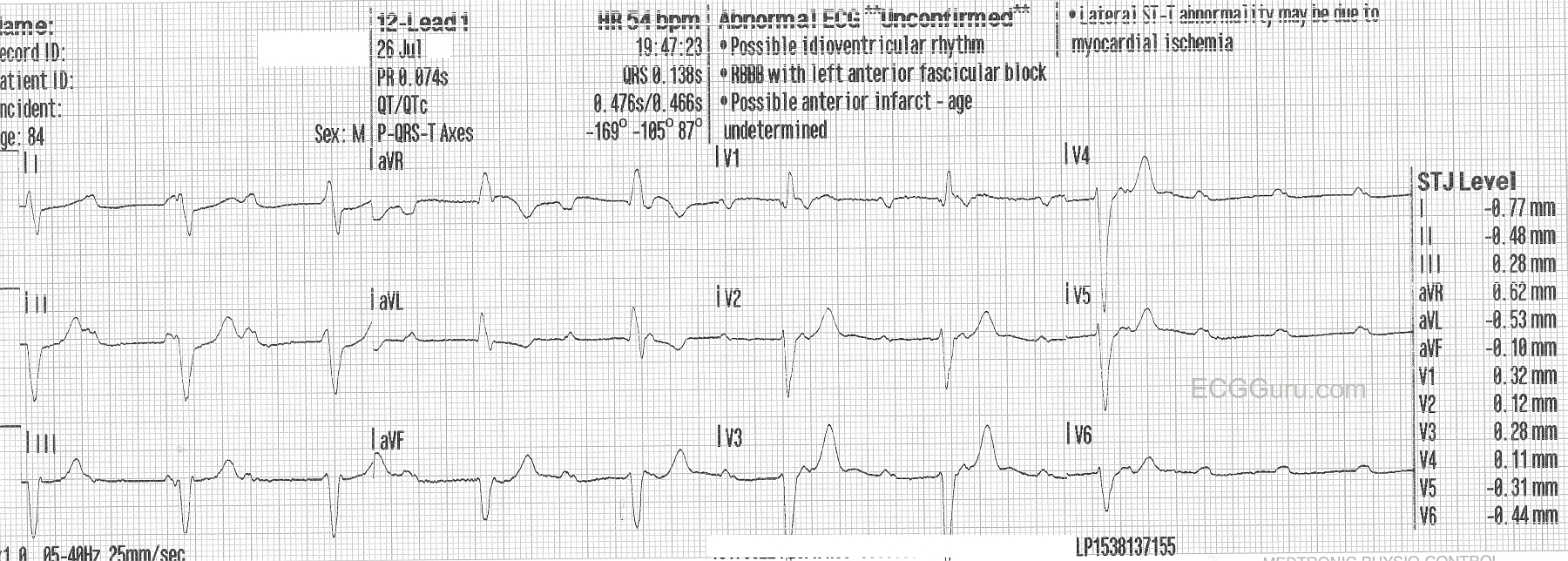
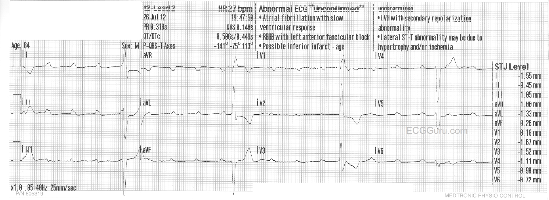
This week's ECG of the WEEK was donated to us by Sebastian Garay. These two ECGs were obtained less than 30 seconds apart from an 84 year-old man who called fire-rescue because he felt dizzy and fell.  He was not injured in the fall, and his vital signs remained stable, with an adequate BP.  These two ECGs were obtained prior to arrival in the Emergency Dept.

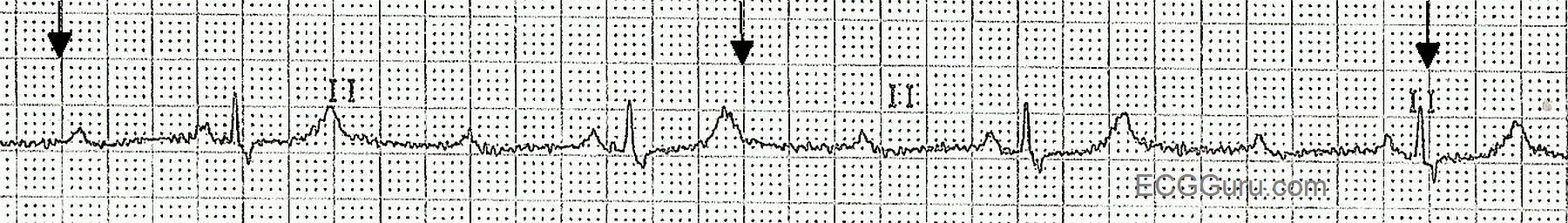
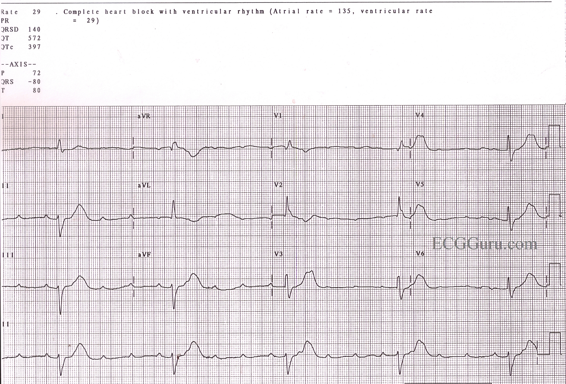
The first one shows a sinus rhythm at about 110/min.  There is a complete heart block (third-degree AV block), and the escape rhythm is a wide-complex rhythm at a rate of about 54/min and slowing severely toward the end.  The second ECG was taken less than 30 seconds after the first, and shows a significantly slower escape rhythm rate at 27/min., while the sinus rate increases to 120/min.  The change is sinus rate is likely an attempt by the nervous system to compensate for the lower cardiac output as the ventricular rate slows. The escape rhythm is not only slower, but there are some changes in the QRS morphology from the first ECG.

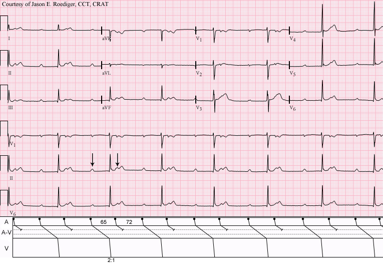
For your basic students, this ECG serves to demonstrate the AV dissociation seen in complete heart block.  It is easy to "march out" the P waves, and see that some of them are "hiding" in the QRS comlexes.  It also shows how quickly a rhythm can change rates.

For your more advanced students, you will want to have a discussion about escape rhythms.  This one initially has a fairly fast rate, suggesting junctional origin. The QRS morphology is of the right bundle branch type, with left anterior fascicular block.  However, ventricular rhythms originating from the posterior fascicle region can have the "RBBB / LAFB" morphology.  If this escape rhythm is fascicular (ventricular) in origin, it is an accelerated idioventricular rhythm.  The second escape rhythm appears very similar to the first, with the very noticeable exceptions of QRS morphology, especially in V1 and V2, and the rate.

This patient was given Atropine in the ED, with no change to the rhythm.  We do not know what transpired after that, but suspect a pacemaker was in his future. 

We look forward to comments from our members about these two very interesting ECGs.


jer5150's picture

LYME DISEASE CAUSING AV BLOCK

We are now in the month of August.

Dawn's picture

Sinus Tachycardia With High-grade AV Block

This ECG has always caused a lively conversation in ECG classes, both beginner classes and advanced. There is an obvious underlying sinus tachycardia, with clear P waves.

Some propose that the ECG shows a second-degree AVB, Type II, in that the PR intervals are constant, or nearly so. There is a slight discrepency if you compare the first PRI with the others. The QRS complexes, while very slow, have a pattern of right bundle branch block with left anterior fascicular block - not an unlikely finding in second-degree AV block, Type II, since that is a block in the fascicles of the interventricular conduction system. Type II blocks usually are accompanied by signs of bundle branch dysfunction.

Others strongly believe this is a third-degree, or complete, heart block. They argue that the PR intervals are not identical, and propose that a longer strip would uncover the discrepency. The wide QRS complexes have a strong left axis deviation, which could support the argument for idioventricular escape rhythm.

What do you think? Please comment below.

Pages

All our content is FREE & COPYRIGHT FREE for non-commercial use

Please be courteous and leave any watermark or author attribution on content you reproduce.