Dawn's picture

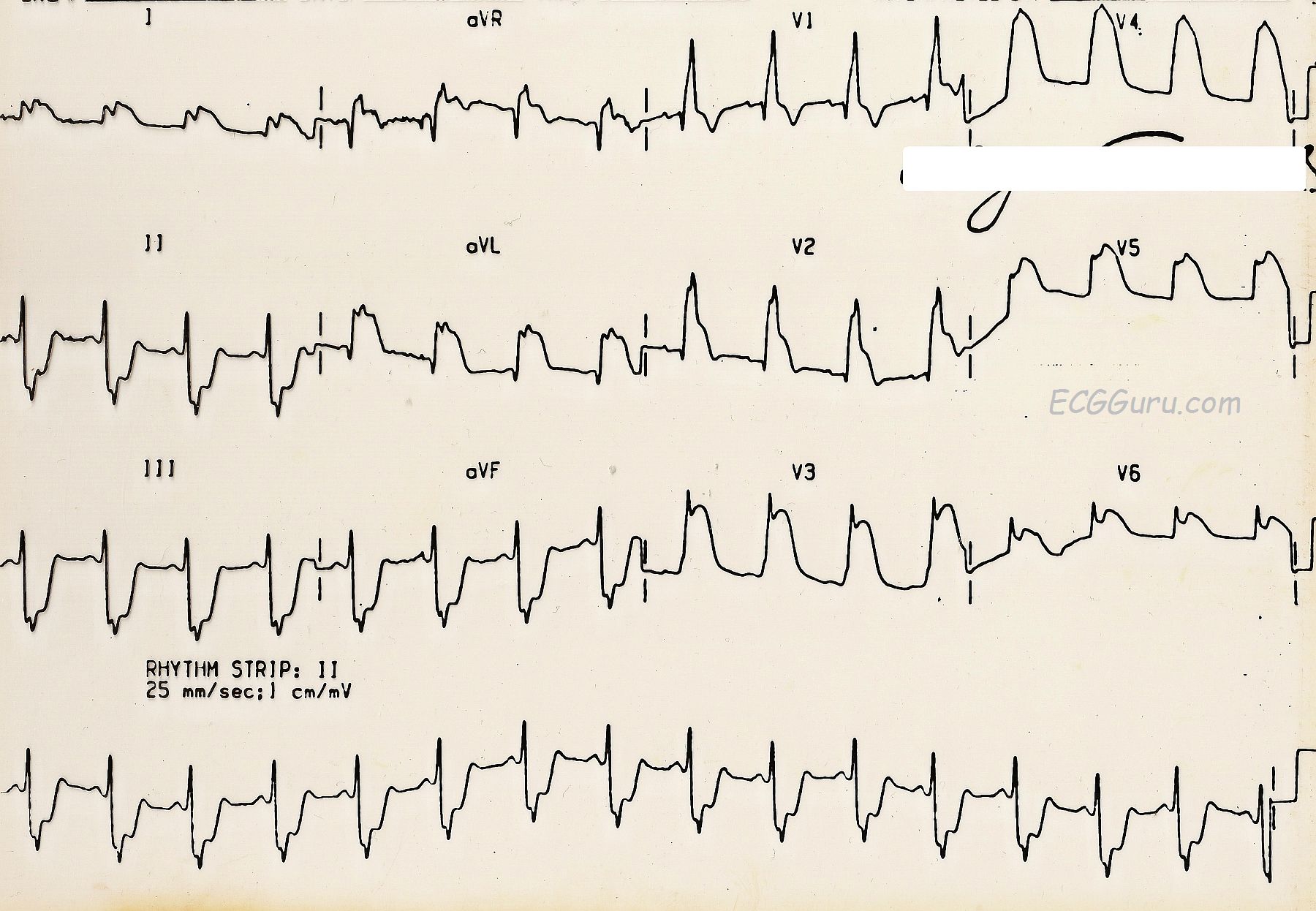
This ECG depicts an extensive and ultimately, fatal, injury.  There is marked ST segment elevation in Leads V2 through V6 (anterior wall).  There is also ST elevation in Leads I and aVL (high lateral wall).  The ST elevation in aVR is indicative of a very proximal lesion in the left coronary artery, which supplies the anterior wall, including the anterior portion of the septum, the high lateral wall, and, in this case, the low lateral wall.  The inferior leads, II, III, and aVF, show reciprocal ST depression.

This is an old ECG - the computer readings of the rate and intervals is lost, as is the grid.  But the rate here appears to be about 80 bpm and the QRS is widened.  There is a right bundle branch block ECG pattern, which is not surprising given the extensive septal damage.  Normally, the criteria for RBBB on the ECG includes an rSR' pattern in V1 (seen here) and a small, wide s wave in Leads I and V6.  This s wave is not seen here, presumably due to the effects of the ST elevation in those leads.

What matters clinically in a patient like this is not whether there is RBBB or another type of interventricular conduction delay. This patient needs immediate restoration of blood flow through the LCA and intensive medical/nursing care.  As mentioned before, this patient did not survive, in spite of being brought to a hospital.  We do not know the exact mechanism of death or treatment course in this case.

If you are teaching students to use multiple leads in assessing rhythm, this is a great example of how one or two leads can be very misleading.  I have used this ECG's V4 in an excercise illustrating this concept.  Shown V4, many people would call this "AIVR" or "V Tach".  Seen in context with the other leads, it is obvious that we are looking at ST elevation that is as high as the R wave.  Two leads are better than one, and twelve are better than two.


Rate this content: 
Average: 5 (2 votes)

Comments

ekgpress@mac.com's picture

     Interesting tracing presented by Dawn this week. As stated  this looks like an "old" ECG  with the grid no longer seen. I'll add a few points to those made by Dawn.
  • I would guess that the heart rate is faster than 80/minute. Obviously difficult to judge given the absence of any grid  but proportionately  the rate looks to be in the 100-110/minute range ( = my guess).
  • The rhythm is sinus. I agree entirely that in the context of acute infarction  the rSR' alone in lead V1 qualifies as "RBBB" ( = Right Bundle Branch Block)  even without the wide terminal S wave that are normally seen in leads I,V6 with complete RBBB.
  • There is also LAHB (Left Anterior HemiBlock) - with a predominantly negative QRS in the inferior leads.
  • Of note  there IS a small initial r wave ( = positive deflection) in lead V1  whereas there is a Q wave (initial negative deflection) in lead V2. In theory  preservation of the initial small positive deflection in lead V1 suggests preservation of septal forces  though given the rest of this ECG, I wouldn't be certain of that presumption ...
  • As noted  there is diffuse and marked ST elevation on this tracing. This is most marked in aVL and V2-thru-V5  but also present in I,aVR,V6. Most patients with acute occlusion of the LMain coronary artery do not live long enough to reach the hospital  but on occasion they may survive long enough for an ECG to be recorded  and this is what I suspect in this case.
  • Distinction between LMain vs proximal LAD (Left Anterior Descending) coronary artery occlusion is often difficult. Statistically (as suggested above LMain occlusion is much less common. That said  factors suggesting LMain occlusion in this case include: i) ST elevation in aVR clearly more than in V1; and ii) Truly diffuse and marked ST elevation with RBBB/LAHB in association with more ST elevation in aVR > aVL.
  • Of note  there is already a large Q in lead aVL  and there is profound reciprocal ST depression in the inferior leads which is as marked as the ST elevation in midprecordial leads is.
  • This patient's unfortunate demise is not an unexpected result.
  • NOTE: The ECG entity of severe coronary disease/possible LMain involvement  should not be confused with this tracing, in which acute LMain occlusion is suspected. With severe LMain disease — there is diffuse ST segment depression (with possible exception of ST elevation in leads aVR, V1). In contrast, with acute LMain occlusion (which is rarely seen) — there will be diffuse ST segment elevation, often accompanied by severe conduction defects. This case probably represens one of those rare instances in which an ECG captured acute evolving LMain occlusion in this patient who ultimately did not survive ...
For those wanting more on determination of the "culprit artery" with acute STEMI  Please check out this pdf (excerpted from my ECG-2014-ePub CLICK HERE TO DOWNLOAD. The part on distinction between LMain vs proximal LAD occlusion is covered in Sections 10.24, 10.25.
  • NOTE: For more on distinction between LMain disease vs LMain occlusion — Please See Section 09.40 in our pdf on Using Lead aVR (from my ECG-2014-ePub).
  • This case is linked to my ECG Blog #103


Ken Grauer, MD  www.kg-ekgpress.com   [email protected] 

All our content is FREE & COPYRIGHT FREE for non-commercial use

Please be courteous and leave any watermark or author attribution on content you reproduce.