Dawn's picture

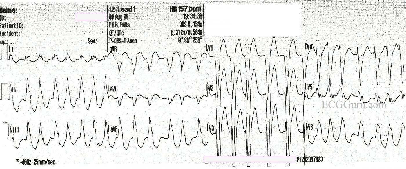
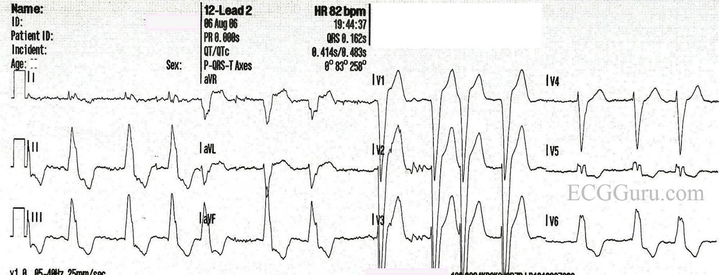
This is a good example of atrial fibrillation with left bundle branch block.  You get two ECGs with this one, because the patient presented to EMS with a fast heart rate, and the rate was slowed with the drug diltiazem.  We do not have any other patient information, unfortunately.

In the first strip, we see a wide-complex tachycardia.  In an emergency situation, with an unstable patient, this rhythm could safely be treated using an emergency ventricular tachycardia (VT) protocol.  In fact, all WCTs should be considered to be VT until proven otherwise.  In most emergency settings, the unstable VT patient would be electrically cardioverted, which will often convert atrial fibrillation as well.  The stable patient with this rhythm would be treated with an antiarrhythmic drug, such as amiodarone.  This may convert or slow down atrial fib.  So, in the initial stages of treatment, differentiating between VT and A Fib is not the first priority.  Assessing the patient's hemodynamic stability and addressing the rate if necessary are the priorities.  

So, how do we know this is NOT VT?  It can be difficult, but in this case, the rhythm, even though fast, is very irregular.   VT is not always perfectly regular, but this irregularly-irregular rhythm points to atrial fibrillation. Also, the pattern of the QRS morphology fits with LBBB.  The criteria for LBBB are:  1) supraventricular rhythm, 2) wide QRS, 3) negative QRS in V1 with positive QRS in Leads I and V6.  If we assume the rhythm is atrial fibrillation, we meet the first criteria.  The other two are self-evident.

After the medication is administered, 10 minutes later, we see the rate slow down.  There is no change in the irregularly-irregular rhythm, and the LBBB pattern remains.  All that has changed is the rate and, hopefully, the patient's symptoms.  This confirms that the original rhythm was not VT.  

Remember, atrial fib lowers cardiac output because there is no P wave - no "atrial kick".  Also, the fast rate associated with new-onset atrial fib often compromises ventricular filling and cardiac output.   LBBB also has a deleterious effect on cardiac output.  Wide QRS complexes indicate that the ventricles are not contracting efficiently and synchronously.  The left ventricle is depolarizing by way of a slow wave of depolarization, rather than all the cells getting the message to depolarize at the same time.  Having these two conditions at the same time can have a very negative effect on cardiac output, leading to CHF.  The first step in treatment often involves simply slowing the rate to normal, which allows for better ventricular filling and decreases the workload on the heart.  Then, the fibrillation and bundle branch block can be addressed.

There is some very slight variation in the QRS morphology in both of these ECGs.  We will leave it to the more advanced Gurus to comment on this, and any other interesting features we may have missed.

Rate this content: 
Average: 2.9 (8 votes)

Comments

ekgpress@mac.com's picture

     Interesting set of tracings by Dawn on a very common problem. I would like to "revisit" the premise of the 1st tracing (= ECG #1) - in which there is a fairly rapid WCT (Wide-Complex Tachycardia) without identifiable P waves. Should this be "assumed VT till proven otherwise and treated according?"

     My ANSWER is: i) It depends; and ii) "Ya gotta be there". Thus, as much as we'd like "the Answer" - there just is NO one single answer for "all comers" to this question with a tracing such as we initially see.
  • TRUE - If the patient is clearly hemodynamically unstable because of the fast rhythm with a tracing like ECG #1 - then electricity (cardioversion) is the treatment of choice. But the reality is - that it is very uncommon (if not rare) that rapid AFib not associated with WPW needs "emergency" cardioversion. Almost always - such patients will be hemodynamically stable enough to allow other treatment measures (ie, of acute heart failure). Treating pulmonary edema does wonders for fast AFib!
  • Therefore - I think a better answer for ECG #1 is a relative probability answer as a working diagnosis. This rhythm is clearly IRREGULAR. Even without calipers there is variation almost from one beat to-the-next. Although VT is not always completely regular - it is rarely as irregularly irregular as we see here. In addition, more just the upright monophasic QRS in leads I,V6 and the negative QRS in lead V1 - the morphology of the QRS in all leads is completely typical of LBBB. Note how steep the downslope of the S waves in V1,V2,V3 is - which is highly characteristic of LBBB. While this doesn't rule out the possibility of VT with 100% certainty (since VT may present with lbbb-like morphology) - it does make VT far less likely. Complete LBBB as seen here is common in patients with significant underlying heart disease - so the scenario of rapid AFib with LBBB morphology is one that is often seen. The history (even with a patient you've never seen before) will often clue you into the likelihood that rather than VT - we are dealing with acute exacerbation (often of heart failure) in a patient with underlying LBBB and AFib.
  • It sounds like the EMS team agreed with my above rationale - as they gave Diltiazem. IF you think the rhythm is ischemic VT - then Diltiazem is absolutely contraindicated - as its vasodilatory and negative inotropic effects typically precipitate deterioration of VT to VFib. But if you assess ECG #1 as rapid AFib with underlying LBBB - then Diltiazem is an agent of choice for controlling the ventricular response.
  • So - While I would NOT be 100% certain of the rhythm diagnosis for ECG #1 the first time I looked at it - I would be >95% comfortable that this is not VT, but rather rapid AFib with underlying LBBB. I would remain ready to cardiovert IF neded - but my hunch would be that the rapid AFib and call to EMS was the result of exacerbation of some underlying disorder (probably heart failure) - and that rate slowing (ie, with Diltiazem) and treatment of the underlying medical condition might be all that is needed to gain control of the situation.
  • As to the slightly variable QRS morphology that Dawn referred to - I don't think we see enough to speculate on what may or may not be a rate-related conduction abnormality vs movement from sick patient. In either case - QRS variation is not enough to alter management considerations.
P.S. IF your patient with ECG #1 truly is hemodynamically unstable because of this fast rhythm - then by all means cardiovert immediately. Please just keep in mind that most of the time the patient will NOT be hemodynamically unstable as to mandate emergency cardioversion when the rhythm is AFib not related to WPW.
 

Ken Grauer, MD  www.kg-ekgpress.com   [email protected] 

All our content is FREE & COPYRIGHT FREE for non-commercial use

Please be courteous and leave any watermark or author attribution on content you reproduce.