Dawn's picture

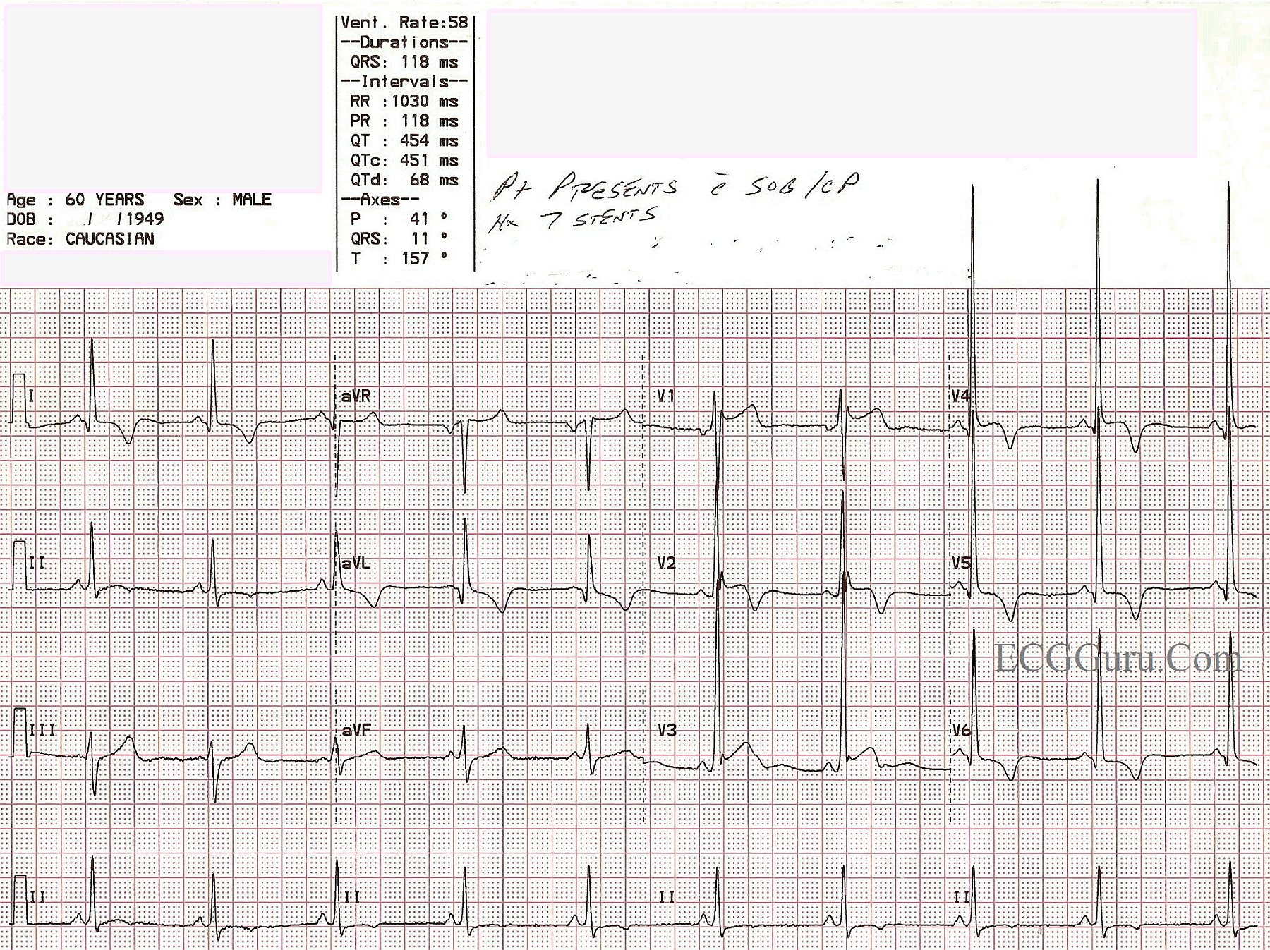
This 60-year-old man presented to the Emergency Department with chest pain and shortness of breath.  He gave a history of having seven coronary artery stents in the past.

This is a good ECG for demonstrating the voltage and ST criteria for LVH and acute anterio-lateral wall M.I. in the same patient, where both conditions have been confirmed by other tests.   If you are teaching the topics of ST elevation M.I., or left ventricular hypertrophy, you will probably have to address the issue that LVH can be considered a "mimic" for STEMI, especially for beginners.  This is because LVH causes ST depression in leads with upright QRS complexes, and reciprocal ST elevation in leads with negative QRS complexes.  This is called ST segment discordance.  The ST changes in LVH are due to the "strain" pattern, indicating strain on the left ventricular myocardium.  It is true that some ST elevation will appear in V1 and V2 in these patients, and can be mistaken for M.I.  In the ECG shown here, the patient has definite ST elevation in leads which would NORMALLY have depression in the LVH strain pattern.  Leads V3 through V6 and I and aVL have ST elevation that is not discordant, and is definitely real. In addition, the ST elevation in V1 and V2 are greater than expected for LVH alone.

The criteria most often used to determine LVH is the Sokolov-Lyon Criteria, (S wave in V1 + R wave in V5 or V6 = 35 mm).  It is best to confirm the LVH with echocardiograhy.

This patient was taken to the cath lab, and the M.I. was confirmed, although his case was lost to followup, and we do not know if he received more angioplasty, coronary artery bypass, or other treatment, or what his condition was post catheterization.

For a similar ECG and discussion on this site, go to this LINK.

As always, the information presented with the Instructors' Collection series is very basic, and we welcome and look forward to comments from the ECG GURUs out there!


Rate this content: 
Average: 4 (3 votes)

Comments

Thanks for EKG, has short PR any relationship here?

ekgpress@mac.com's picture

 
This tracing just looks wierd! I completely agree with Dawn given the history from this 60yo man with new-onset chest pain and dyspnea - that ACS (Acute Coronary Syndrome) is clearly suggested. Lateral precordial lead R wave amplitude is dramatically increased, virtually guaranteeing true left ventricular chamber enlargement. In addition:
  • There are Q waves in lateral leads I, aVL; V4,V5,V6 - with the Q in lead aVL clearly deeper and wider than should be expected for septal q waves.
  • ST segments are elevated and/or coved in multiple leads.
  • T wave inversion appears more symmetric in lateral leads than would be expected for simple "strain" (LVH repolarization changes).
That said - there are a number of unusual aspects of this tracing worthy of comment:
  • Transition occurs early (between V1-to-V2) - with result that the R wave is already 20 mm tall by V2. This is distinctly unusual for simple LVH ...
  • There is an rSr' complex in lead V1 - with saddleback ST elevation in this lead. This pattern is consistent with a Brugada Type II ST segment - that while definitely not diagnostic of Brugada syndrome, nevertheless brings up latent Brugada syndrome as a possibility (Click HERE for review of Brugada patterns). 
  • There is J-point notching of ST segments in not only lead V1, but also V2,V3. Associated with this is an upwardly concave elevated ST segment that is sometimes seen with early repolarization variants.
BOTTOM LINE: Nothing beats clinical correlation! We learned that this patient has a history of multiple coronary stents in the past - and acute MI was confirmed in the cath lab in association with this tracing. Learning this only piques my curiosity more to wonder what his pre-event ECG would have looked like - but unfortunately, this is no longer available. The lesson to be learned, is that regardless how tempting in might be to ascribe the changes on this tracing to a combination of marked LVH, early transition and early repolarization pattern - given the history of multiple prior stents in this 60yo man with new-onset chest pain - acute ongoing MI has to be the working diagnosis until proven otherwise. Fascinating tracing!
------------------
P.S. To Marionurse (who asked about the PR interval) - I printed out this ECG for optimal magnification. While I completely agree that the PR interval "looks short" - I measure it out to be 0.12 second, which is the lower limit of normal. I do not see any deflections consistent with delta waves. Review of prior tracings would clearly help - but based on this single ECG, I'd interpret the PR interval within the normal range. 
  • Much has been made in the past about "LGL" (Lown-Ganong-Levine) - for when the PR looks short but there is no delta wave. Realize that 2 reasons other than existence of an accessory pathway for the PR to be short are: i) enhanced conduction; and ii) smaller anatomic AV node size. Sometimes I acknowledge a short PR interval (ie, of 0.10 or 0.11 second in an adult) - by writing, "short PR" on my interpretation. In the absence of a history of cardiac arrhythmias - the isolated finding of minimal PR interval shortening in an adult (esp. a 60 year old man) is almost negligible. Likelihood of a potentially important underlying AP (Accessory Pathway) remains low unless the PR is <0.10 second. The measured PR = 0.12 second in this case is within the lower normal range.

Ken Grauer, MD  www.kg-ekgpress.com   [email protected] 

All our content is FREE & COPYRIGHT FREE for non-commercial use

Please be courteous and leave any watermark or author attribution on content you reproduce.