
Submitted by Dr A Röschl on Sun, 09/10/2023 - 07:42
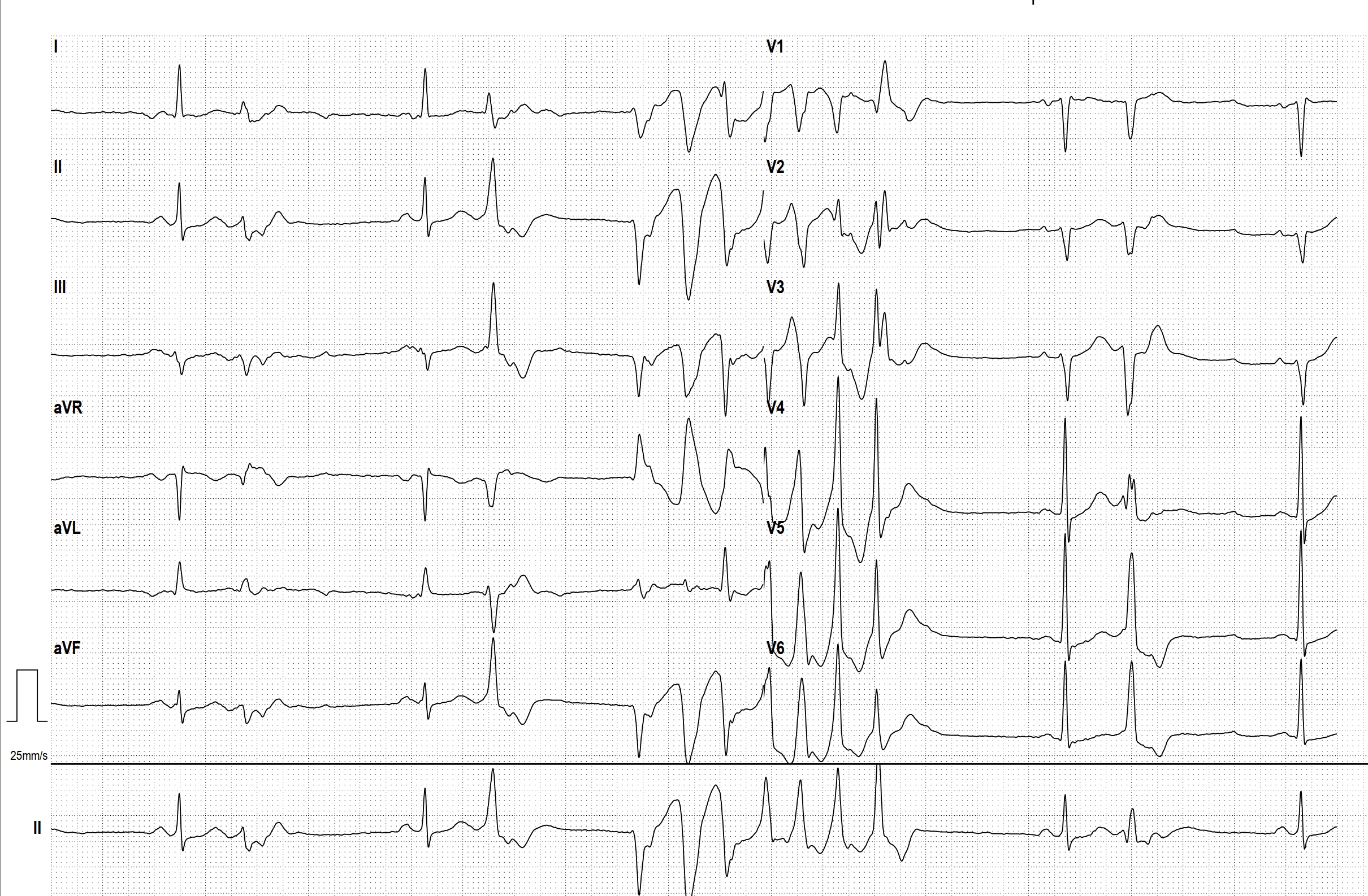
This EKG shows a sinus rhythm with ventricular bigeminy and retrograde conduction leading to retrograde depolarization of the sinus node, resulting in a longer pause (sinus node reset).
Then, a polymorphic ventricular tachycardia occurs over 7 beats. The QT interval of the sinus beats does not appear prolonged, thus ruling out Torsades de Pointes tachycardia. The most likely cause of this type of polymorphic ventricular tachycardia during a stress EKG is cardiac ischemia/coronary artery disease.
Rate this content:
-

- Dr A Röschl's blog
- Log in or register to post comments
All our content is FREE & COPYRIGHT FREE for non-commercial use
Please be courteous and leave any watermark or author attribution on content you reproduce.
Comments
Coupling Interval versus Presence of Prolonged QT Interval
Thanks for the very informative rhythm strips. These are great! I feature two chapters on this topic in my new book, "Getting Acquainted With Wide Complex Tachycardias - A Workbook for the Electrocardiographically Confused!".
In cases of polymorphic ventricular tachycardia - which includes torsade de pointes (TdP) and non-torsade polymorphic VT - the couplng interval between the last sinus beat and the ectopic beat that precipitates the polymorphic VT is often a better determinant of TdP or non-torsade polymorphic VT than the presence of a prolonged QT interval.
A coupling interval > 400 msec is most likely TdP (and its coupling intervals are usually much, much greater than 400 msec) while a coupling interval = or < 400 msec is most likely a non-torsade polymorphic VT (which is the case with this rhythm strip). And - as you indicated - the most common cause of a non-torsade polymorphic VT is ischemia. Although there is a long pause between the PVC and the actual onset of the polymorphic VT here, it isn't neceassry for the onset of a non-torsade polymorphic VT because the non-torsade polymorphic VTs are not pause-dependent.
With monomorphic VTs, we look at the ectopic rhythm to learn more about the origin of the tachydysrhythmia. With polymorphic VTs, we have to look at a bit of sinus rhythm to learn more about the origin. It is especially helpful to actually see the onset of the tachycardia (as you have demonstrated here) when differentiating between TdP and non-torsade polymorphic VT. Fortunately, that is not too difficult because both types of polymorphic VTs tend to be very paroxysmal, giving us the ability to see a little bit of sinus rhythm plus the onset of another episode of tachycardia.
Diagnosing "torsade de pointes" based on the iconic, spindle-shaped ECG pattern with the "twisting of the pointes" is also error-prone because the non-torsade polymorphic VTs can, at times, present with the same pattern. And torsade de pointes does not always show a spindle-shaped pattern - especially with shorter episodes.
Thanks so much for the valuable addition of your ECGs and rhythm strips along with your excellent discussions on this website. It's very much appreciated by all of us who study ECGs and who come to this website for more information on electrocardiography!
Jerry W. Jones MD FACEP FAAEM
https://www.medicusofhouston.com
Twitter: @jwjmd