
Submitted by jer5150 on Sat, 09/29/2012 - 10:17
This is another one of those instances where I fortuituously viewed this ECG the day it was performed in our triage department several years ago. I immediately knew what was happening here but apparently nobody in the emergency department saw the obvious clues.
Patient's clinical data: 53-year-old white man with long 37-year history of tobacco abuse, hypothyroidism, and COPD. Patient stopped smoking 4 weeks prior to current admission. No definite fever. Had some intermittent CP. Lung nodules were identified in last 2 weeks and patient was scheduled for biopsy. Presented to the ED with SOB worsened X 5 days, coughing, sore throat, and difficulty swallowing. The ED physician's provisional diagnosis for this patient was pneumonia and subsequently the patient was given his first dose of antibiotics prior to his admission upstairs to the cardiac step-down ward.
Vital signs:
Temp: 98-degrees F
Pulse: 138/min
Resp: 22
BP: 131/88
Alert & oriented X 4
Question:
(1.) What is causing the pattern seen in this ECG?
Rate this content:
-

- jer5150's blog
- Log in or register to post comments
All our content is FREE & COPYRIGHT FREE for non-commercial use
Please be courteous and leave any watermark or author attribution on content you reproduce.
Comments
Pericardial effusion causing
Pericardial effusion causing eletrical alternans?
INTERPRETATION
INTERPRETATION:
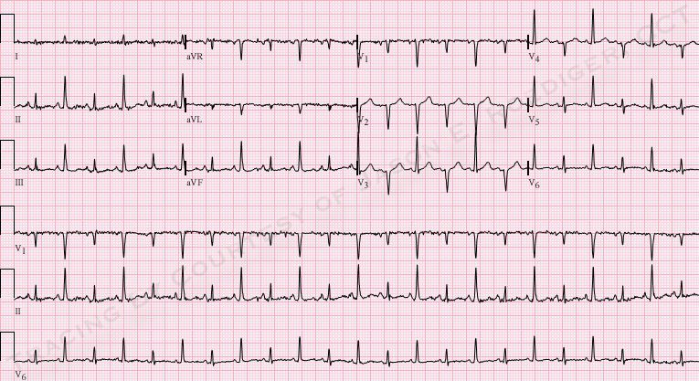
(1.) Sinus tachycardia (rate = 140/min) with . . .
(2.) . . . electrical alternans and . . .
(3.) . . . low voltage QRS suggestive of . . .
(4.) . . . pericardial effusion.
COMMENTS:
This patient had a very large pericardial effusion with tamponade and underwent an urgent pericardiocenesis. He was later diagnosed with stage 4 lung CA and subsequently died about 20 months later.
Jason E. Roediger - Certified Cardiographic Technician (CCT)
[email protected]